Showing posts with label Salk Institute for Biological Studies. Show all posts

Wednesday, September 02, 2026

Are predatory brain immune cells eating nerve cells in Amyotrophic lateral sclerosis (ALS)?

Good news!

"Highlights
  • Salk scientists discover that central nervous system-resident immune cells called microglia use TAM receptors (discovered by study senior author Greg Lemke in 1991) to eat specific nerve cells in mouse model of ALS
  • The study is the first to show TAM receptors can be used to kill living nerve cells
  • The insights contribute to ongoing efforts to harness TAM receptors in clinical settings to treat cancers, autoimmune disorders, and more
...

Neurons are often the center of attention in brain and neurodegeneration research, but other cells in the central nervous system are important, too. Recent studies have shown microglia, which are immune cells specific to the brain and spinal cord, are very active during human ALS. The question is: Why? ...

Active microglia can be easily identified in research by assessing their expression of TAM receptors. ... discovered this family of proteins, which are critical bridges between the immune system and the rest of the body, more than three decades ago. ...

To determine whether the TAM system was the link between microglia activation and motor neuron death in ALS, the ... researchers started with the most widely used mouse model of ALS, called SOD1. These mice express a mutant SOD1 protein that causes ALS in people.

They first found that many motor neurons had been eaten in the spinal cords of SOD1 mice. The levels of TAM proteins were also elevated in these mice, particularly Axl and Mer.
Looking closer at the motor neurons, the scientists noticed those “eat me” signs, which are little molecules called phosphatidylserine, were being displayed on cells when they shouldn’t be. From there, the TAM system springs into action, guiding microglia to their next meal: a live neuron.

The natural next question ... is “what happens when we eliminate Axl and Mer?” Without these two TAM family proteins, the mice got sicker faster but lived longer. ...

“When we looked at how many motor neurons mice without Axl and Mer had, compared to mice with Axl and Mer, we found losing the TAM proteins meant preserving muscle controls.” ..."

From the abstract:
"Activation of microglia is a prominent feature of amyotrophic lateral sclerosis (ALS), a neurodegenerative disease that leads to the death of motor neurons.
A key component of this activation is elevated expression of the TAM receptor tyrosine kinases Axl and Mer (gene name Mertk).
Here we show that germline and microglial-restricted inactivation of the Axl and Mertk genes in the SOD1G93A mouse model of ALS leads to an extension of lifespan, which is tied to the preservation of cholinergic motor neurons and neuromuscular synapses.
Also elevated on SOD1G93A neuronal surfaces is the essential TAM co-ligand phosphatidylserine, a potent ‘eat-me’ signal through which apoptotic cells are engulfed by microglia.
Correspondingly, we find that microglial lysosomes are filled with the remains of cholinergic neurons in the SOD1G93A spinal cord, whereas this accumulation is markedly reduced in the SOD1G93AAxl-/-Mertk-/- cord.
Together, these results suggest that microglia phagocytically kill living neurons, and thereby hasten death in ALS."

Are predatory brain immune cells eating nerve cells in ALS? – Salk Institute "Salk Institute scientists discover central nervous system immune cells find and kill specific nerve cells in the spinal cords of mice with late-stage ALS, exacerbating the condition; findings could be harnessed in cancer and autoimmune therapy innovation efforts"


Microglia (green) contain the remnants of motor neurons (purple) inside microglial lysosomes (yellow).


Tuesday, March 31, 2026

Why does the body deem some foods safe and others unsafe?

Amazing stuff!

"Highlights
  • Scientists identify three new proteins, one each from soybean, corn, and wheat, that the body uses to determine oral tolerance—the opposite of food allergy
  • They found that specialized immune cells called regulatory T cells interact with these proteins in the gut
  • By understanding tolerance, researchers can better understand food allergies and begin to imagine future immunotherapies that promote tolerance rather than allergic reactions
...

identifies new bits of food proteins that tell gut immune cells when to tolerate certain foods. They found three of these protein segments, called epitopes—one each from soybean, corn, and wheat. These epitopes interact with specialized immune cells called regulatory T cells to inform that tolerance-or-rejection decision. ..."

"Editor’s summary
Immunological tolerance to dietary antigens is essential for preventing food allergies and digestive disorders such as celiac disease. However, the specific food-derived antigens that contribute to immune tolerance remain poorly described. Blum et al. mapped the dietary epitopes recognized by food-responsive T cell receptors (TCRs) derived from murine intestinal regulatory T (Treg) cells.
Seed storage proteins from corn, wheat, and soy, including the maize protein αZein, were targets of food-responsive Treg cell TCRs. αZein-specific Treg cells suppressed T cell responses to αZein ex vivo and after adoptive transfer into naive mice. These findings provide insight into the dietary components recognized by naturally occurring Treg cells that mediate oral tolerance. ...

Abstract
Food antigens elicit immune tolerance through the action of intestinal regulatory T (Treg) cells. Unlike food allergens, the proteins that mediate tolerance are mostly undescribed.
Here, we found that epitopes derived from seed storage proteins are targets of murine intestinal Treg cells, with the most frequent response targeting the C terminus of the maize protein alpha-zein.
A major histocompatibility complex (MHC) tetramer loaded with this antigen revealed that zein-specific T cells are predominantly intestinal Treg cells, develop concurrently with weaning, and constitute up to 2% of the peripheral Treg cell pool. Zein-responsive Treg cells repressed naïve T cell proliferation ex vivo, and prior dietary exposure resulted in a constrained response upon diverse inflammatory challenges in vivo, supporting a specific role for gut-resident Treg cells in suppressing systemic immune responses.
Our work reveals the development, immune-suppressive characteristics, and function of naturally occurring Treg cells that recognize dietary seed storage proteins, a previously undescribed class of antigens in oral tolerance."

Why does the body deem some foods safe and others unsafe? - Salk Institute for Biological Studies "Study co-led by scientist now at Salk Institute finds three new proteins that the body uses to determine “safe” foods, helping understand food tolerance and allergy"

What changes happen in the aging brain? A comprehensive single-cell atlas to date of epigenetic changes in the aging mouse brain

Very impressive!

"Highlights
  • Salk researchers create epigenetic atlas of cell type-specific changes in the aging mouse brain
  • The atlas represents eight different brain regions and 36 different cell types, and shows clear epigenetic differences associated with different ages
  • The new resource—available publicly on Amazon Web services—can be used to unravel age-related contributions to neurodegenerative diseases like Alzheimer’s, Parkinson’s, and ALS
...

One major mechanistic influence on aging is epigenetic change: the way small chemical tags on top of our base genetic code shift over time to alter gene expression. Salk researchers created the most comprehensive single-cell atlas to date of epigenetic changes in the aging mouse brain, revealing how DNA methylation, genome structure, and gene activity change across brain regions and cell types. The new atlas represents eight brain regions and 36 distinct brain cell types, with over 200,000 single cells profiled across methylation and chromatin conformation assays, plus nearly 900,000 cells captured with spatial transcriptomics. ..."

What changes happen in the aging brain? - Salk Institute for Biological Studies "Salk Institute scientists create atlas of cell type-specific epigenetic changes that occur in the mouse brain as it ages to better understand the basis of neurodegenerative diseases"


Salk researchers used spatial transcriptomics to map where different cell types reside in the mouse brain. Shown are excitatory neurons (left, blue), inhibitory neurons (middle, red), and non-neuronal cells (right, green), color-coded by cell type.


Sunday, December 21, 2025

Astrocytes stabilize circuits in the adult mouse brain using the protein CCN1, which could be targeted to restore plasticity in injury, disorder, or disease

Good news! Amazing stuff! Could be a breakthrough!

"... This flexibility is called neuroplasticity, and our ability to learn, make new memories, and recover from injury all depend on it. ... team have now discovered a molecule that is critical for stabilizing brain circuits in adulthood: a protein called CCN1 secreted by star-shaped cells called astrocytes. The CCN1 pathway could now be a prime target for new therapeutics designed to support learning and plasticity in conditions like Alzheimer’s disease, depression, or PTSD, or to promote neural repair after injury or stroke."

From the abstract:
"Neural circuits in many brain regions are refined by experience. Sensory circuits support higher plasticity at younger ages during critical periods—times of circuit refinement and maturation—and limit plasticity in adulthood for circuit stability. How astrocytes, a glial subtype, maintain these differing plasticity levels, and whether they stabilize the properties of sensory circuits in adulthood, remain largely unclear.
Here we take a comprehensive approach to address these questions and establish astrocytes as key orchestrators of circuit stability.
Combining a transcriptomic approach with ex vivo electrophysiology and in vivo imaging, we identify that astrocytes release CCN1 to maintain synapse and circuit stability in the adult visual cortex.
Overexpressing CCN1 in astrocytes during the critical period promotes the maturation of inhibitory neurons, limits ocular dominance plasticity and promotes oligodendrocyte differentiation and maturation.
Conversely, knocking out astrocyte CCN1 in adults destabilizes binocular circuits and reduces myelination.
This establishes CCN1 as an astrocyte-secreted factor that stabilizes neuronal circuits by coordinating the maturation state of multiple cell types, and demonstrates that the composition and properties of sensory circuits require ongoing maintenance in adulthood, and that these maintenance cues are provided by astrocytes."

🧠 SALK DECEMBER NEWS 🧠


Fig. 1: Identifying CCN1 as an astrocyte pro-stability factor in the mouse visual cortex.


Friday, September 26, 2025

How does the brain differentiate painful from non-painful touch?

Amazing stuff! Good news! Makes sense! 😊

"After nine months in the womb, humans enter a world filled with texture and shape. We must then quickly learn to recognize and respond to textures and objects in the outside world, beginning with sensations like the soft feel of a t-shirt or the doughy squish of a sandwich. By learning what touch sensations are innocuous, the brain can better recognize painful insults that might cause damage—think skinning a knee or stubbing a toe. But 7 to 10 percent of the global population develops mechanical allodynia, a form of chronic pain where innocuous light touch is perceived as painful. ...

neuroscientists have discovered that altered neuronal activity in a brain area called the dorsal column nuclei drives mechanical allodynia. The dorsal column nuclei process and categorize light touch information before sending it on to cortical areas that discriminate between different types of touch. In monitoring the activity of mouse neurons in the gracile nucleus, they discovered that mechanical allodynia causes the gracile nucleus’ normal neural activity patterns to become uncoordinated. ...

The dorsal column nuclei’s representation of touch gets transmitted to another area called the thalamus, which also plays a prominent role in pain perception. However, this is mediated by different thalamic regions that receive noxious pain information from specialized pain receptors termed nociceptors.
Normally, these two pathways remain separated, but in the 
case of mechanical allodynia, it seems that their wires get crossed at the level of the thalamus and above, in the cortex. Understanding how these wires get crossed is the next step in determining how the brain distinguishes innocuous from painful touch. ...

“By looking at both inflammation-induced and injury-induced mechanical allodynia, we were able to search for common signatures in the patterns of neural activity that characterize this form of chronic pain,” ... “This led us to observe changes in neural processing that point to the dorsal column nuclei as crucial in discriminating innocuous touch from potentially harmful touch.” ..."

From the highlights and summary:
"Highlights
• The DCN is a critical node for mechanical allodynia but not thermal allodynia
• Mechanical allodynia disrupts neural population activity and neural correlations in the GrN
• Altered GrN neural correlations predict increased information transmission to the VPL
• GrNSst VPL projection neurons are required for the expression of mechanical allodynia

Summary
The neural circuits that transmit the sense of pain and how pain is encoded by these circuits are still poorly understood. 
Mechanical allodynia is a prominent form of chronic pain characterized by painful responses to innocuous touch that develops as a consequence of nerve damage and inflammation.
Here, we show that alterations to the normal log-normal distribution of neuronal activity and structure of neural correlations between neurons in the dorsal column nuclei (DCN) constitute a signature feature of mechanical allodynia, with the transmission of “allodynic” light touch information to the thalamus by somatostatin-positive projection neurons in the DCN being essential for its expression and development.
Our findings are consistent with neural activity in the DCN encoding a reference landscape of tactile percepts that animals use to differentiate innocuous touch from potentially noxious mechanical stimuli."

How does the brain differentiate painful from non-painful touch? - Salk Institute for Biological Studies "Salk scientists use mouse model to pinpoint gracile nucleus as brain area responsible for discriminating between painful and non-painful touch, with its dysfunction leading to chronic pain"


Graphical abstract


If your last name is Virlogeux like Amandine Virlogeux (first author of the study from Fance, sounds to me a bit like virologist), what are your prospects to become involved in biological studies like at the Salk Institute? Just kidding! (this is the best photo Google search was able to find of her. The Salk Institute itself does not feature a better photo (tres dommage)).


Saturday, August 30, 2025

Scientists debut a new foundational atlas of the plant life cycle

Good news!

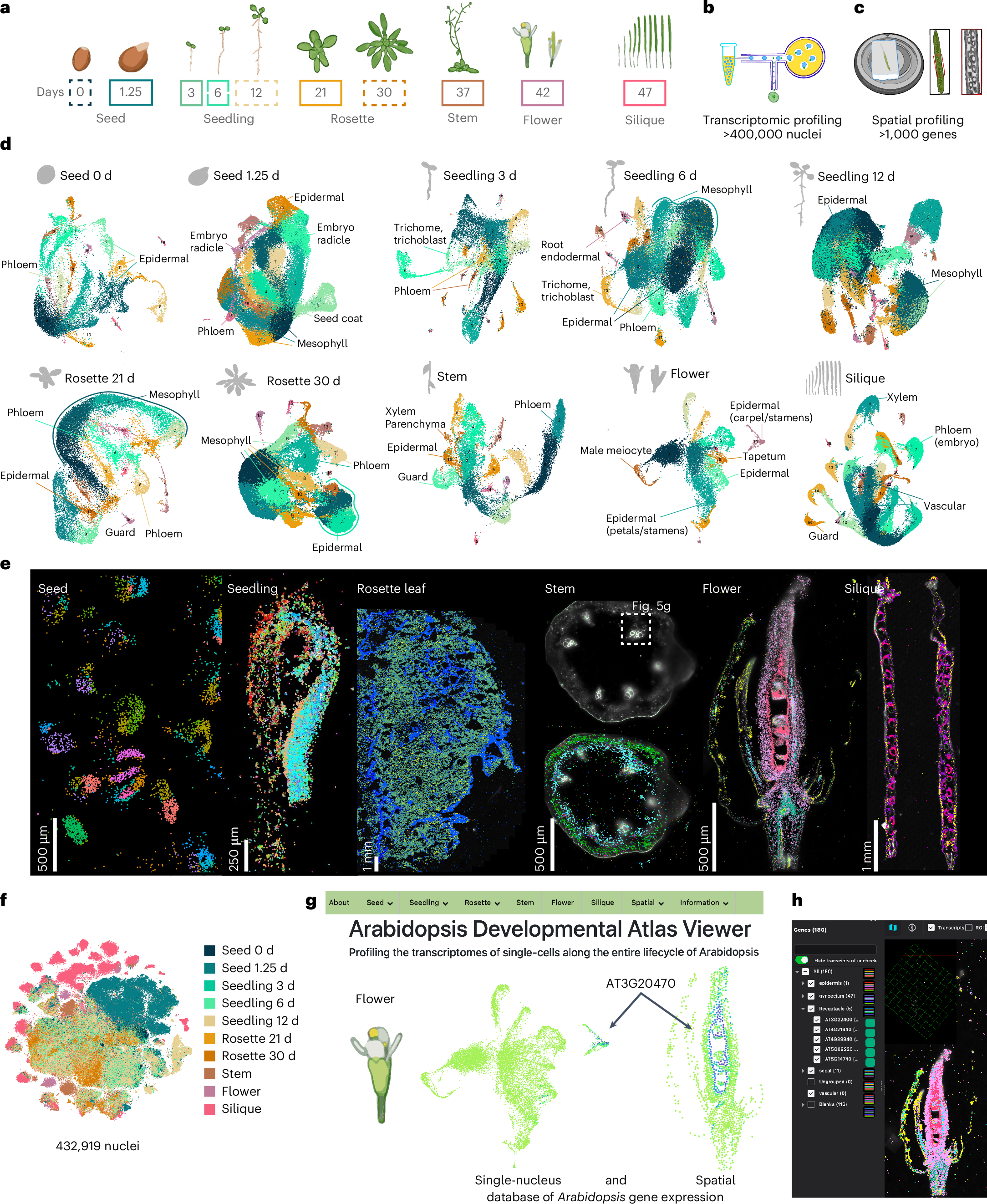
"... researchers have now established the first genetic atlas to span the entire Arabidopsis life cycle. The new atlas—created using detailed single-cell and spatial transcriptomics—captures the gene expression patterns of 400,000 cells within multiple developmental stages as Arabidopsis grows from a single seed to a mature plant. The publicly available resource will be hugely informative to future studies of different plant cell types and developmental stages, and how they respond to stress and environmental stimuli. ..."

From the abstract:
"Arabidopsis has been pivotal in uncovering fundamental principles of plant biology, yet a comprehensive, high-resolution understanding of its cellular identities throughout the entire life cycle remains incomplete.
Here we present a single-nucleus and spatial transcriptomic atlas spanning ten developmental stages, encompassing over 400,000 nuclei from all organ systems and tissues—from seeds to developing siliques. Leveraging paired single-nucleus and spatial transcriptomic datasets, we annotate 75% of identified cell clusters, revealing striking molecular diversity in cell types and states across development.
Our integrated approach identified conserved transcriptional signatures among recurrent cell types, organ-specific heterogeneity and previously uncharacterized cell-type-specific markers validated spatially.
Moreover, we uncover dynamic transcriptional programs governing secondary metabolite production and differential growth patterns, exemplified by detailed spatial profiling of the compact yet complex apical hook structure; this profiling revealed transient cellular states linked to developmental progression and hormonal regulation, highlighting the hidden complexity underlying plant morphogenesis.
Functional validation of genes uniquely expressed within specific cell contexts confirmed their essential developmental roles, underscoring the predictive power of our atlas.
Collectively, this comprehensive resource provides an invaluable foundation for exploring cellular differentiation, environmental responses and genetic perturbations at high resolution, advancing our understanding of plant biology."

Scientists debut a new foundational atlas of the plant life cycle - Salk Institute for Biological Studies "Salk Institute researchers map every cell type and developmental state across the entire life cycle of model plant Arabidopsis"


Fig. 1: A spatially resolved transcriptional atlas of the Arabidopsis life cycle.


Scientists arrive at minimal, viable plant genomes through large chromosomal deletions

Amazing stuff!

"... Ancient events in plant evolution have left behind large, duplicated regions in their genomes.

... scientists found that deleting these large blocks of DNA can still lead to normal plants.

The findings demonstrate that large chromosomal deletions are a viable strategy in plant genetic engineering, which could now accelerate the development of streamlined, minimal plant genomes—a major goal in industries looking to create new plant-based biotechnologies. ...

The researchers used CRISPR-Cas9 to delete four large, duplicated blocks in Arabidopsis thaliana, a model plant commonly used in plant biology research. The deletions were then verified using whole-genome sequencing, which revealed minimal off-target effects. ..."

From the significance and abstract:
"Significance
Plant genomes are shaped by ancient polyploidy events, leaving behind extensive duplicated regions whose biological roles remain largely uncharacterized.
In this study, we demonstrate that targeted deletion of large, retained syntenic blocks in Arabidopsis thaliana results in viable plants, some with distinct phenotypes and widespread transcriptomic changes.
These findings challenge the assumption that such regions are essential and highlight the potential redundancy or modularity within plant genomes. Our approach of removing entire duplicated blocks offers a powerful strategy to functionally dissect conserved genomic regions, investigate gene linkage and dosage effects, and accelerate the development of streamlined, minimal plant genomes. This work establishes a scalable framework for genome engineering with broad implications for plant biology, synthetic genomics, and biotechnology.

Abstract
Plant genomes have undergone multiple rounds of whole-genome duplication (WGD) throughout their evolutionary history. As a result, many species, including Arabidopsis thaliana, retain duplicated genomic segments, or syntenic regions, which harbor large numbers of paralogous genes preserved from these ancient WGD events.
We deleted four large, duplicated blocks, ranging from ~115 kb to ~684 kb using Staphylococcus aureus Cas9 to explore the effects of knocking out these blocks in Arabidopsis.
Large deletions like these remain rare, especially in small and gene-dense plant genomes. Deletions were subsequently verified using whole-genome sequencing, which revealed minimal off-target effects.
The number of deleted genes ranged from 16 to 60, and transposable elements ranged from 4 to 112 among the four deleted blocks.
Two deletion lines showed distinct phenotypes resulting from the loss of many genes, while two others displayed no obvious defects, including for flowering time or hypocotyl elongation.
Moreover, RNA-sequencing revealed that expression compensation, where deletions of paralogous genes lead to the upregulation of intact paralogues, was not a general response to the deleted regions under the conditions tested.
Thus, it is possible to obtain viable plants when deleting large fragments that may be redundant or that contain nonessential genes.
These results demonstrate that large chromosomal deletions can be used as a tool for genome engineering approaches, such as genome minimization in plants and allele replacement using homology-directed repair and other precision editing methods. Targeted deletions of large chromosome fragments will be a valuable tool for research and biotechnology applications."

Scientists get back to basics with minimal plant genomes | EurekAlert!


Fig. 4 Generation of deletion lines (deletions of retained duplicate regions or nonlethal genomic segments) and their applications. 


Thursday, March 28, 2024

Modeling the origins of life: New evidence for an “RNA World”

Amazing stuff!

"... New research ... now provides fresh insights on the origins of life, presenting compelling evidence supporting the RNA World hypothesis. ... unveils an RNA enzyme that can make accurate copies of other functional RNA strands, while also allowing new variants of the molecule to emerge over time. These remarkable capabilities suggest the earliest forms of evolution may have occurred on a molecular scale in RNA. ..."

From the significance and abstract:
"Significance
An RNA enzyme with RNA polymerase activity was used to replicate and evolve an RNA enzyme with RNA-cleavage activity. The fidelity of the polymerase is sufficient to maintain heritable information over the course of evolution, with a succession of variants of the RNA-cleaving RNA enzyme arising that have progressively increasing fitness. The RNA-catalyzed evolution of functional RNAs is thought to have been central to the early history of life on Earth and to the possibility of constructing RNA-based life in the laboratory.
Abstract
An RNA polymerase ribozyme that was obtained by directed evolution can propagate a functional RNA through repeated rounds of replication and selection, thereby enabling Darwinian evolution. Earlier versions of the polymerase did not have sufficient copying fidelity to propagate functional information, but a new variant with improved fidelity can replicate the hammerhead ribozyme through reciprocal synthesis of both the hammerhead and its complement, with the products then being selected for RNA-cleavage activity. Two evolutionary lineages were carried out in parallel, using either the prior low-fidelity or the newer high-fidelity polymerase. The former lineage quickly lost hammerhead functionality as the population diverged toward random sequences, whereas the latter evolved new hammerhead variants with improved fitness compared to the starting RNA. The increase in fitness was attributable to specific mutations that improved the replicability of the hammerhead, counterbalanced by a small decrease in hammerhead activity. Deep sequencing analysis was used to follow the course of evolution, revealing the emergence of a succession of variants that progressively diverged from the starting hammerhead as fitness increased. This study demonstrates the critical importance of replication fidelity for maintaining heritable information in an RNA-based evolving system, such as is thought to have existed during the early history of life on Earth. Attempts to recreate RNA-based life in the laboratory must achieve further improvements in replication fidelity to enable the fully autonomous Darwinian evolution of RNA enzymes as complex as the polymerase itself."

Modeling the origins of life: New evidence for an “RNA World” - Salk Institute for Biological Studies Salk scientists unveil RNA capabilities that enable Darwinian evolution at a molecular scale, and bring researchers closer to producing autonomous RNA life in the laboratory

Sunday, July 11, 2021

How neurons get past “no”

Recommendable! About neuronal threshold modulation!

"... The study, published May 25, 2021, in the journal Cell Reports, shows that inhibitory neurons do more than just inhibit neuron activity like an off-switch; paradoxically, they actually increase the amount of information transmitted through the nervous system when it needs to be flexible. To make this possible, inhibitory neurons need to be integrated into the circuit in a specific way. These observations could help scientists better understand and treat disorders involving our ability to focus and modulate signals based on the bigger picture, which are altered in conditions such as anxiety and attention deficit disorders. ...
While the researchers tested the theory in neurons involved in vision, the findings could apply widely to neurons found throughout the brain and nervous system ..."

"... We find that modulation is less detrimental than other forms of neuronal variability and that its negative effects can be nearly completely eliminated if modulation is applied selectively to sparsely responding neurons in a circuit by inhibitory neurons. We verify these predictions in the retina where we find that inhibitory amacrine cells selectively deliver modulation signals to sparsely responding ganglion cell types. Our findings elucidate the central role that inhibitory neurons play in maximizing information transmission under modulation."

How neurons get past “no” - Salk Institute for Biological Studies

Tuesday, May 04, 2021

How brain cells repair their DNA reveals “hot spots” of aging and disease

Good news! Possibly a breakthrough! This is probably some critical research!

"... Now, a new study by Salk scientists finds that these repairs are not random, but instead focus on protecting certain genetic “hot spots” that appear to play a critical role in neural identity and function. ...
“This research shows for the first time that there are sections of genome that neurons prioritize when it comes to repair,” ... “We’re excited about the potential of these findings to change the way we view many age-related diseases of the nervous system and potentially explore DNA repair as a therapeutic approach.” ...
The authors found approximately 65,000 hot spots that covered around 2 percent of the neuronal genome. They then used proteomics approaches to detect what proteins were found at these hot spots ...
Previous research has focused on identifying the sections of DNA that suffer genetic damage, but this is the first time researchers have looked for where the genome is being heavily repaired. ..."

"Humans have only a limited capacity to generate new neurons. These cells thus need to repair errors in the genome. To better understand this process, [researchers] developed Repair-seq, a method to locate DNA repair within the genome of stem cell–derived neurons. DNA repair hotspots (DRHs) were more likely to occur within specific genomic features such as gene bodies as well as in genomic formations, open chromatin, and active regulatory regions. This method showed that repair was enriched at sites involved in neuronal function and identity. Furthermore, proteomic data indicated that genes in DRHs are enriched in Alzheimer's disease and that DRHs are more active in aging. ..."

"Neurons are the longest-lived cells in our bodies and lack DNA replication, which makes them reliant on a limited repertoire of DNA repair mechanisms to maintain genome fidelity. These repair mechanisms decline with age, but we have limited knowledge of how genome instability emerges and what strategies neurons and other long-lived cells may have evolved to protect their genomes over the human life span. A targeted sequencing approach in human embryonic stem cell–induced neurons shows that, in neurons, DNA repair is enriched at well-defined hotspots that protect essential genes. These hotspots are enriched with histone H2A isoforms and RNA binding proteins and are associated with evolutionarily conserved elements of the human genome. These findings provide a basis for understanding genome integrity as it relates to aging and disease in the nervous system."

How brain cells repair their DNA reveals “hot spots” of aging and disease - Salk Institute for Biological Studies Salk scientists reveal new insights into neurodegenerative disorders and potential for genetic therapies

Here is the link to the underlying research article:

Monday, March 01, 2021

Never-before-seen antibody binding revealed, informing both liver cancer and antibody design

Good news! Perhaps a breakthrough for better treatments! Cancer is history! Like Covid-19 and HIV/AIDS before, cancer will be defeated! It is only a question of time!

"In structural biology, some molecules are so unusual they can only be captured with a unique set of tools. That’s precisely how a multi-institutional research team led by Salk scientists defined how antibodies can recognize a compound called phosphohistidine—a highly unstable molecule that has been found to play a central role in some forms of cancer, such as liver and breast cancer and neuroblastoma. ...
Previously, ... showed that phosphorylation on the amino acid tyrosine can drive cancer progression, a discovery that led to numerous anticancer drugs. More recently, Hunter turned his attention to phosphorylation of the amino acid histidine (which creates phosphohistidine), suspecting that the process might also play a role in human disease. ...
... developed a suite of antibodies able to bind to phosphohistidine in proteins, and used chemically stabilized phosphohistidine analogues to develop a series of monoclonal antibodies that could recognize these forms. The next step was to understand exactly how the antibodies are able to bind to phosphohistidine. This led ... to collaborate with ... a world-renowned expert in using protein crystallography to define antibody structures, to study the structures of the phosphohistidine antibodies.
“... have been collaborating on this project for the past seven years,” ... “We have obtained new insights into how antibodies can evolve to recognize phosphates linked to proteins, which is very satisfying.”
To find out how phosphohistidine is recognized, they needed to image their antibodies in the act of binding the phosphohistidine, and so formed crystals between each antibody bound to a phosphohistidine peptide. ...
The two types of antibody crystal structures solved by the team revealed exactly how different amino acids are arranged around the phosphohistidine to bind it tightly. Their five structures more than double the number of phospho-specific antibody structures previously reported, and provide insights into how antibodies recognize both the phosphate and the linked histidine. They also reveal at a structural level how the two types of antibody recognize different forms of phosphohistidine, and this will allow the scientists to engineer improved antibodies in the future.​ ..."

"Phosphohistidine (pHis) is a labile posttranslational modification with two isoforms, 1-pHis and 3-pHis, involved in many cellular processes across the kingdoms of life. Due to its lability, it is difficult to study the pHis modification using standard biochemical tools and techniques. ..."

Salk team reveals never-before-seen antibody binding, informing both liver cancer and antibody design - Salk Institute for Biological Studies Multi-institute collaboration uses X-ray crystallography and recombinant antibodies to uncover workings of an elusive molecule central to human health

Here is the link to the referenced paper:

Sunday, February 28, 2021

Specific bacteria in the gut prompt mother mice to neglect their pups

Amazing stuff! We still know so little about the multitude of bacteria living in our gut and their effects on behavior and the brain!

"As scientists learn more about the microorganisms that colonize the body—collectively called the microbiota—one area of intense interest is the effect that these microbes can have on the brain. A new study led by Salk Institute scientists has identified a strain of E. coli bacteria that, when living in the guts of female mice, causes them to neglect their offspring. ... 
“To our knowledge, this is the first demonstration that the intestinal microbiota is important for promoting healthy maternal behavior and bonding between mom and offspring in an animal model,” ...
“It adds to the ever-growing evidence that there’s a gut-brain connection, and that microbes are important for regulating the behavior of the host that they’re inhabiting.” ..."

"Maternal behavior is necessary for optimal development and growth of offspring. The intestinal microbiota has emerged as a critical regulator of growth and development in the early postnatal period life. Here, we describe the identification of an intestinal Escherichia coli strain that is pathogenic to the maternal-offspring system during the early postnatal stage of life and results in growth stunting of the offspring. ..."

Specific bacteria in the gut prompt mother mice to neglect their pups - Salk Institute for Biological Studies Salk researchers make unexpected discoveries about how a particular microbe influences animal behavior

Here is the link to the referenced paper:

Sunday, November 03, 2019

Mysterious microproteins have major implications for human disease

Very recommendable! Microproteins (up to 100 amino acids) could become a new, very promising research area! 


Article also describes how difficult it was to tag the microprotein PIGBOS with a florescent protein. The researchers discovered "As they mapped PIGBOS’ location ... sits on the outer membrane of the mitochondria ... They were surprised to see PIGBOS interacting with a protein called CLCC1, which is part of an organelle called the endoplasmic reticulum (ER). ... The researchers found that PIGBOS actually communicates with CLCC1 to regulate stress in the ER. Without PIGBOS, the ER is more likely to experience stress, which leads to a chain of events where the cell tries to clear out harmful misshapen proteins (called the unfolded protein response). If the cell fails to dispose of these proteins, it will initiate a self-destruct sequence and die." 

Those kind of interactions between cell organelles have rarely been studied before.


Mysterious microproteins have major implications for human disease - Salk Institute for Biological Studies: LA JOLLA—As the tools to study biology improve, researchers are beginning to uncover details into microproteins, small components that appear to be key to some cellular processes, including those involved with cancer. Proteins are made up of chains of linked amino acids and the average human protein contains around 300 amino acids. Meanwhile, microproteins have fewer than 100 amino acids.