Showing posts with label virology. Show all posts

Friday, July 31, 2026

Ancient DNA confirms historical accounts of how smallpox got to the Americas. Really!

Again the AAAS gets caught spreading stereotypes or biases!

By repeating a narrative it does not become a truth! Or as Oscar Wilde once said: "The truth is rarely pure and never simple" quoted from Oscar Wilde's 1895 play The Importance of Earnest.

It also appears that the researchers were biased. Why would they enter the year 1492 CE as the start dated for the sequenced ancient genomes in their abstract (see below)? The arrival of Christopher Columbus in the Americas? Is this a pretense of exactness or demagoguery?

This research is based only on two individuals found in one cemetery in Chile! This study also involves some serious estimation work on the evolution of the virus. Was this properly validated?

Maybe the Europeans of 16th and 17th century did not think that smallpox was so contagious anymore. Very likely, these Europeans did not understand that the indigenous population might be more vulnerable to this virus.

What diseases were spread from indigenous people to the early arriving Europeans?
"The primary and practically sole major disease widely accepted by historians and scientists to have passed from the Americas (including South America) back to early European arrivals is syphilis (specifically non-venereal or venereal treponemal variants that adapted into the European syphilis epidemic of the 1490s)." (Google Search)
Syphilis in the 16th century was significantly severe, disfiguring, and frequently deadly. (Google Search)

Or why did not other, more contagious diseases evolve in South America or the American continent before the arrival of Europeans?

"Sometime around the turn of the 17th century, two people died in northern Chile. Their disease-ravaged bodies were bundled in layers of textiles, buried in an Indigenous cemetery, and naturally mummified by the desert environment. Now, scientists have identified their killer: smallpox. The virus’ genome found in their bones and described today in Science represents the first molecular evidence of the disease in the colonial Americas, confirming historical accounts of the disease arriving with European contact and eventually killing millions of Indigenous inhabitants. ...

surprising hints that the smallpox virus’ evolution might have slowed down between the 16th and 18th centuries ...

the Chilean strain differed from modern smallpox, but thinks it was likely less virulent. ...

Archaeologists had previously described some of the mummies as having skin lesions, which they attributed to arsenic exposure. But smallpox could also leave such lesions. ...

Evolutionarily, the Chilean strain sits between pox viruses circulating during the Viking age and a virus isolated by Poinar’s team from a Lithuanian child mummy who died between 1643 and 1665 C.E. It likely diverged from other smallpox lineages around 1296 C.E., and went extinct before the early 20th century.
By estimating how long it would take for the strain’s hallmark genetic variants to evolve and combining that with radiocarbon dates from one of the mummy’s hairs, the scientists deduced that the outbreak in northern Chile occurred between 1492 and 1631 C.E."

"... Smallpox was one of the deadliest diseases introduced to the Americas during European colonization. The rapid spread of the disease across North, Central, and South America is estimated to have resulted in the deaths of three to four million people, particularly among Indigenous populations with no prior immunity. ...

Bruno González and colleagues identified ancient smallpox virus DNA in two mummified Indigenous Inca-Early Colonial-era individuals recovered from northern Chile, who lived between 1492-1631 CE."

Caveat: I did not read the research article.

From the editor's summary and abstract:
"Editor’s summary
Smallpox was introduced to the Caribbean by European colonialists before quickly spreading south. Romero González et al. sequenced ancient smallpox genomes from two likely Incan individuals from Chile dated between about 1492 and 1631 CE. Skin lesions on these mummified remains were previously attributed to arsenic exposure, but they are also consistent with smallpox infection.
Phylogenetic analyses suggested that this smallpox strain was most closely related to ancient European lineages that went extinct before the 20th century. These represent the earliest smallpox genomes found in the Americas and help set a timeline for gene loss during the evolution of this deadly virus. ...

Abstract
Smallpox was a major driver of population collapse in the Americas after European contact, yet the genetic identity of the causal strains remains unknown.
Here, we report the first ancient smallpox genomes in the Americas, dating to approximately 1492–1631 common era (CE), recovered from two Inca-Colonial individuals in northern Chile.
These genomes form a now-extinct lineage that diverged around 1296 CE, after the splitting of early medieval European strains but before the emergence of modern variola lineages, providing direct molecular evidence for smallpox introduction through European colonization.
We further identify a constant tempo of gene inactivation until the late 16th century, followed by a phase of constraint and a subsequent rebound in substitution rates, linking variola virus evolution to major shifts in human demography and epidemiology."

Ancient DNA confirms historical accounts of how smallpox got to the Americas Science | AAAS "“Landmark” paper finds disease originated with European contact"

Sunday, July 12, 2026

Covid-19 Pandemic recreated in Fast Forward in a test tube

Good news! However, how the SARS-CoV-1 virus was released from the Institute of Virology in Wuhan (a dual use research institute at the time), China will not be answered, I suspect.

"A key step in the origin of many pandemics occurs when an animal-borne virus infects humans and then evolves to spread more efficiently from person to person. That is why scientists and physicians keep a close watch on viruses that could jump from animals to humans, such as emerging strains of avian flu and bat coronaviruses, as well as viruses that have already crossed into humans but, for now, spread poorly among people, such as hantavirus and Ebola.

Researchers have now recreated in a test tube, within just a few months, the evolutionary path the coronavirus followed during the COVID-19 pandemic – from the original Wuhan strain to the emergence of the highly contagious Omicron variants. ..."

From the abstract:
"In vitro protein evolution can provide powerful insights into the amino acid sequences that underlie key biological functions.
Here, we use this to explore the evolutionary trajectories of the SARS-CoV-2 spike protein receptor-binding motif (RBM) binding the human angiotensin-converting enzyme 2 (ACE2), an essential first step in viral infection.
Applying stringent selection pressures starting from the Wuhan or another non-Omicron variant protein-coding sequence results in rapid convergence towards Omicron characteristic mutations and its sub-lineages.
Conversely, under mild selection, only some Omicron-like mutations are selected, however at lower frequencies and with incomplete representation.
Stringent selection results in fewer, but dominant, non-synonymous mutations mirroring Omicron mutations and their variations within its sub-lineages.
Notably, initiating evolution from Omicron itself results in maintenance of Omicron-defining mutations under both conditions.
This evolutionary pattern parallels global SARS-CoV-2 mutation trends as well as in silico simulations, emphasizing the critical role of receptor-binding constraints in shaping viral adaptation.
Mutations primarily associated with immune evasion are not selected by in vitro evolution.
Our findings demonstrate the predictive capacity of in vitro evolution, suggesting Omicron RBM to be the humanized binding motif, emerging from high-stringency selection, superimposed on milder background pressures."

Pandemic in Fast Forward - Life Sciences | Weizmann Wonder Wander - News, Features and Discoveries "Israeli and Czech scientists recreated the coronavirus’s evolutionary journey in a test tube – revealing the conditions that can produce highly contagious variants"


Fig. 1: High-throughput yeast display evolution of SARS-CoV-2 RBD under defined selection pressures.


Fig. 3: Mutation accumulation following in vitro evolution in comparison to SARS-CoV-2.


Tuesday, July 07, 2026

Mucosal immunoglobulin A (IgA) to prevent norovirus infection in mice

Good news!

"... The findings ... upend traditional vaccine development efforts which to date have focused on creating norovirus-fighting antibodies that circulate in the blood rather than taking up residence in the gut.

According to findings, mucosal IgA (Immunoglobulin A) antibodies — which are found primarily in the body’s mucosal surfaces, including the lungs, airway, the intestines, and gut — provide key immune defense against norovirus. ...

“By using mouse models, we discovered that IgA was both necessary and sufficient for protection against norovirus.” ..."

From the editor's summary and abstract:
"Editor’s summary
Although most individuals recover from norovirus infection after an unpleasant few days, some individuals, such as infants, older adults, and immunocompromised individuals, are at risk of developing severe gastroenteritis. As such, vaccines to combat this infection are urgently needed. Here, Ökten et al. investigated which components of the immune system are required to effectively clear norovirus in a murine model. The authors found that intestinal IgA in particular was both necessary and sufficient for clearance of norovirus in mice.
To highlight the translational potential of these findings, the authors showed that delivery of an mRNA encoding an anti-norovirus dimeric IgA was able to protect mice.
Together, these results highlight the requirement for a successful norovirus vaccine to specifically elicit mucosal IgA and suggest that anti-norovirus IgA antibodies may offer a prophylactic or therapeutic option for at-risk populations. ...

Abstract
Human norovirus is the leading cause of viral gastroenteritis, yet effective vaccines and therapeutics remain elusive. Using murine norovirus as a model, we found that mucosal immunoglobulin A (IgA) is both necessary and sufficient for protection against infection, whereas CD8+ T cells are dispensable.
Robust intestinal IgA production requires at least 4 weeks of enteric infection, consistent with kinetics of human norovirus RNA clearance.
Systemic vaccination elicits high titers of neutralizing serum IgG but fails to prevent enteric norovirus infection, phenocopying a recent human norovirus vaccine failure.
In contrast, prophylactic delivery of dimeric anti-norovirus IgA via mRNA lipid nanoparticles confers sterilizing immunity.
Together, these findings define a critical role for mucosal IgA in norovirus protection and identify IgA-based treatments as a therapeutic approach for human norovirus."

Gut punch: New study knocks out norovirus where it strikes | Yale News "Gut-homing antibodies provide powerful protection against norovirus, a new Yale study finds, revealing a potential new path for developing vaccines and therapies."

Saturday, June 06, 2026

'World-first' vaccine designed by artificial intelligence to provide broad protection from thousands of variants of viruses

Amazing stuff!

"Artificial intelligence has been used to develop a "fundamentally new" type of vaccine that could protect against large swathes of viruses and prevent pandemics, say researchers.

The team at the University of Cambridge say it is the first time a vaccine's key component has been designed entirely by AI and then trialled in people.

The vaccine was engineered to work on all coronaviruses which would include all Covid variants and viruses that infect animals, but could start the next pandemic. ..."

"The first human clinical trial of a universal Sarbeco coronavirus vaccine, developed by the University of Cambridge and spin-out DIOSynVax (DVX) Ltd, has shown that the vaccine is safe and has no significant side-effects. ..."

From the highlights and abstract:
"Highlights
• pEVAC-PS is a novel, needle-free DNA vaccine targeting all sarbecoviruses
• pEVAC-PS was safe and well tolerated in this phase I study
• Followup Phase 2 to provide data on the breadth and durability of immunogenicity to pEVAC-PS

Summary
Background
Coronaviruses such as SARS, SARS-CoV-2 and related Sarbeco-Coronaviruses continue to pose global health threats, underscoring the need for vaccines capable of inducing broad cross-sarbecovirus protection.
The pEVAC-PS vaccine was developed using Digitally Immune Optimised Synthetic Vaccine (DIOSynVax) technology and pre-clinically selected for the ability to induce broadly protective immune responses across the Sarbecoviruses including SARS, SARS-CoV-2, and related viruses representing potential zoonotic spillovers. For this first-in-human study, the antigen was delivered as a DNA vaccine to enable thermostability and needle-free intradermal administration to support future deployment in resource-limited settings.

Methods
This open label phase I dose escalation study investigated the safety, tolerability and immunogenicity of the pEVAC-PS vaccine candidate against SARS, SARS-CoV-2 and related Sarbeco Coronaviruses via needle-free intra-dermal delivery using the PharmaJet Tropis Device. ...

Findings
Between December 2021 and September 2023, a total of 39 volunteers were vaccinated. The vaccine was well tolerated at all four doses with no significant safety concerns elicited. Interpretation of immunogenicity outcomes was influenced by high baseline antibody levels and heterogeneous exposure histories due to ongoing waves of Omicron variant infections during recruitment, which differed across dose-escalation cohorts and introduced unavoidable immune bias.

Interpretation
Needle-free intradermal delivery of this novel computationally designed PanSarbeco vaccine was safe and well tolerated. Although immunogenicity was modest in the context of substantial pre-existing immunity, participants developed measurable responses to conserved, vaccine-encoded sarbecovirus epitopes, supporting the feasibility of this antigen design strategy."

'World-first' vaccine designed by artificial intelligence

New ‘universal vaccine’ technology could protect us from future virus outbreaks (original news release) "A Cambridge-led team has developed a way to engineer better vaccines that could provide broad protection from thousands of variants of viruses - such as coronaviruses or Ebola - in a single vaccine. This represents a fundamental new vaccine technology that could prevent future pandemics before they begin."


Fig. 2 Vaccination and SARS-CoV-2 variant timeline. The timeline plot shows the vaccinations periods for each group in relation to the dominant SARS-CoV-2 variant circulating in the UK at the time.


Sunday, May 24, 2026

Andes hantavirus PCR test created in two days

Good news! Impressive! This is only the beginning!

"Researchers have managed to develop a diagnostic PCR test for the Andes hantavirus in just two days. The disease sickened more than 10 people and killed three aboard the cruise ship MV Hondius last month. A PCR test is important to track the spread of the virus, which can take up to 42 days to become symptomatic. At the moment, the US Centers for Disease Control and Prevention is using a blood test to detect antibodies in infected people, but it doesn’t work in those not yet showing symptoms."

"... Some other countries have used PCR tests to detect hantavirus, but in the U.S., the Centers for Disease Control and Prevention does not yet have a validated one for diagnosis. The CDC is developing such a test, but in the meantime, the agency has been using a blood test that can detect antibodies in infected people who are symptomatic, but it can’t detect low levels of the virus in asymptomatic people. ..."

P.S. I was unable to find an original news release for this story.

Nature Briefing: Translational Research

Inside the race to develop a hantavirus PCR test "Researchers at the Nebraska Public Health Laboratory worked round the clock to develop a test for the Andes virus at the center of the deadly cruise ship outbreak"

Saturday, May 09, 2026

Satellite virus spreads through viral Trojan Horses

Amazing stuff!

"Satellite viruses replicate their genomes within host cells but depend on helper viruses for spread.
Deltavirus, or hepatitis D-like virus, is a hepatitis B satellite virus that causes severe viral hepatitis in humans.
Recently, deltaviruses have been found in many animals outside of the liver, suggesting that their diversity and disease potential are underestimated.
The current paradigm for satellite viruses is that they simply “borrow” envelope proteins from related helper viruses.
McKellar et al. used electron and super-resolution microscopy in rhabdovirus, herpesvirus, and arenavirus systems to show that deltavirus ribonucleoproteins can package themselves within a variety of helper virions. This viral Trojan horse mode of transmission could broaden deltavirus host range and explain overlooked infections in humans."

From the highlights and abstract:
"Highlights
Deltaviruses hitchhike within helper virus virions, using them as viral Trojan Horses
• The Trojan Horse model is mandatory for productive herpesvirus-deltavirus associations
• This mode of propagation favors deltavirus infectivity
• Trojan Horse model warrants screening for extra-hepatic deltavirus infections in humans

Summary
Hepatitis D-like satellite viruses, known as deltaviruses, have been recently discovered in a wide range of animals. These viruses are thought to expropriate glycoproteins from helper viruses to form infectious particles.
Here, we challenge this paradigm and demonstrate that deltaviruses are packaged within helper virus particles, using them as viral Trojan Horses for cell entry. By leveraging orthogonal electron and optical super-resolution microscopy, we visualize deltaviruses enclosed within virions from rhabdo-, herpes-, and arenavirus families.
We show that this conserved hitchhiking mechanism ensures concomitant deltavirus-helper virus spread, thereby promoting the dissemination of deltaviruses, broadening their host range, and expanding their tropism.
Our findings reveal a previously unrecognized mode of viral transmission, providing a framework to investigate overlooked deltavirus infections outside of the human liver."

In Other Journals | Science


Graphical abstract


Figure 1 A subset of VSV virions is morphologically modified after superinfection of deltavirus-replicating cells


Tuesday, April 21, 2026

Ten different animal species feast on bats in a Ugandan cave offering clues to how deadly viruses spread

Serious stuff!

"When researchers in Uganda set up camera traps to monitor African leopards (Panthera pardus pardus) and spotted hyenas (Crocuta crocuta) in a national park last year, they had no idea that they would record so much more than just those animals. Several of the traps, placed outside a cave known to host Egyptian fruit bats (Rousettus aegyptiacus), caught on video a multitude of creatures feasting on the winged mammals. The bats are known carriers of Marburg virus, which can transfer into humans and cause a fatal haemorrhagic fever, so the footage offers real-time insight into how disease can spread.

Scientists know that bats can transmit viruses to humans either directly, or through an intermediate animal, from forensic detective work and other studies. The team in Uganda thinks this is the first time that potential intermediate animals have been caught on camera in a known hotspot for Marburg virus, which is in the same family as Ebola virus. ..."

‘Bat feast’ animal videos at African cave offer clues to how deadly viruses spread (partially behind paywall) "Researchers filmed 10 species eating or scavenging bats at known Marburg-virus hotspot — and caught hundreds of humans visiting."

Saturday, April 18, 2026

The Ancient Weapons Active in Your Immune System Today

Recommendable!

Caveat: I did not read the entire, long article!

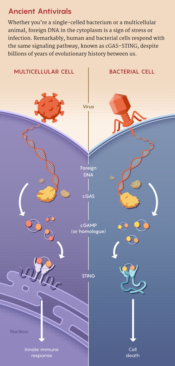
"... In recent years researchers have come upon a surprising finding: Some of the machinery that bacteria use to defend against phages exists, almost unchanged, in our own cells. According to dozens of discoveries made over the past decade, the rules of engagement between cells and viruses were written billions of years ago and still largely define how our innate immune system, the first responder to infection, defends us against viruses and bacteria today. ...

Two recent waves of discovery broke this field open.
First, in 2018, researchers reported a variety of novel bacterial defense systems against viruses(opens a new tab), which now number in the hundreds.
The second wave, starting around 2019, showed that some of these bacterial mechanisms exist in plant and animal cells, including our own — and that they still work the same way they did in those distant ancestors. ...

These and other discoveries that followed reveal an unexplored landscape of human innate immunity — one that could lead to new medical treatments and biotechnological tools ...

A few years later, ... team observed that big constellations of immune genes, including restriction-modification enzymes and CRISPR arrays, tended to cluster together in the same region of bacterial genomes. He and other labs observed that genes of unknown function within these “defense islands” or “genomic islands” could potentially represent novel anti-phage mechanisms. ...

In 2018, his team showed that many of the unknown genes in these defense islands did, in fact, function as a variety of anti-phage defense systems. ..."

The Ancient Weapons Active in Your Immune System Today | Quanta Magazine "Dozens of new discoveries reveal that defenses evolved by bacteria and viruses billions of years ago still define our own innate immune system."


Remarkably, the core machinery of the STING protein (top, protein diagrams) has remained structurally preserved across diverse organisms, although the underlying gene sequence differs widely. Some parts of the protein (bottom, dashed outline) have changed over billions of years.


Monday, April 13, 2026

Slice and dice, a newly characterized biological defense system, directly protects bacteria by chopping up invading viral DNA

Amazing stuff!

"... Bacteria and the viruses that infect them, bacteriophages — phages for short — are ceaselessly at odds, with bacteria developing methods to protect themselves against phages that are constantly striving to overcome those safeguards.

New research ... describes a defense system that is integrated into the protective membrane that encapsulates bacteria. SNIPE, which stands for surface-associated nuclease inhibiting phage entry, contains a nuclease domain that cleaves genetic material, chopping the invading phage genome into harmless fragments before it can appropriate the host’s molecular machinery to make more phages. ..."

From the abstract:
"From mammals to bacteria, the direct recognition and cleavage of viral nucleic acids is a potent defence strategy against viral infection, but it requires mechanisms for distinguishing self from non-self.
In bacteria, CRISPR–Cas and restriction-modification systems achieve this discrimination by recognizing specific DNA sequences or DNA modifications, respectively. Alternative mechanisms probably remain to be discovered.
Here, we characterize SNIPE, an anti-bacteriophage defence system that constitutively localizes to the bacterial cell membrane in Escherichia coli to block phage λ infection.
Using radiolabelled phage DNA and time-lapse microscopy to track phage genomes, we demonstrate that SNIPE directly cleaves phage DNA during genome injection.
Based on proximity labelling, we find that SNIPE associates with host proteins essential for λ genome entry and with the λ tape measure protein, which facilitates λ genome injection across the inner membrane. SNIPE also defends against diverse siphoviruses, probably through direct interactions with their tape measure proteins. Our findings establish SNIPE as a widespread bacterial defence system that exploits the spatial organization of phage genome injection to specifically target viral DNA, representing a previously unknown strategy for distinguishing self from non-self in prokaryotic immune systems."

Slice and dice | MIT News | Massachusetts Institute of Technology "SNIPE, a newly characterized biological defense system, directly protects bacteria by chopping up invading viral DNA."


Fig. 1: SNIPE is a membrane-bound nuclease that provides direct defence against phage.


Fig. 2: SNIPE cleaves phage DNA during genome injection.


Tuesday, March 31, 2026

When phages of different species communicate with each other

Amazing stuff!

"... scientists studied chemical communication by phages (viruses that infect bacteria).

The phages assessed in this study have two choices when they enter a cell: lie dormant or kill the cell and release new virus particles to infect other cells nearby.

It was recently discovered that some phages use chemical communication systems to optimise this decision.

The new study reveals these signals do not pass solely between phages of the same species. Instead, other species – some of them barely related to the signaller – can eavesdrop. ...

The signal chemicals are called peptides, and are produced by the phage during infection. High peptide concentrations signal a lack of susceptible hosts, while low concentrations signal an abundance of uninfected hosts.

The existence of these signalling systems (called “arbitrium” systems) suggests they provide an evolutionary benefit – at least for in-species communication.

But the new study shows “cross-talk” between species does not help the “listener”. ..."

From the highlights and abstract:
"Highlights
• Phages are exposed to non-cognate arbitrium signals from other phages
• Some non-cognate signals mediate crosstalk between phages
• Crosstalk shifts lysis-lysogeny decisions toward early lysogeny
• Crosstalking signals can benefit emitting phages but impose costs on responders

Summary
Many viruses can switch between lytic replication and dormancy (or lysogeny). It was recently discovered that some viruses that infect bacteria (known as bacteriophage or phage) employ peptide-based (“arbitrium”) communication systems to optimize their lysis/lysogeny switch; high peptide concentrations signal a lack of susceptible hosts and trigger lysogeny, while low peptide concentrations signal an abundance of uninfected hosts and prompt lysis.
Here, we demonstrate that arbitrium phages belonging to different species and genera can influence each other’s infection dynamics by secreting similar communication peptides, leading to early lysogenization of the signal-receiving phage and elevated fitness of the signal-emitting phage. Antagonistic coevolution between signal-emitting and signal-receiving phages to manipulate each other’s infection behaviors may explain the rapid diversification of arbitrium systems and their frequent horizontal exchange to escape the noise of crosstalk."

Viruses 'eavesdrop' on each other—but it can backfire


Graphical abstract

Figure 1 Arbitrium phages encounter each other in nature


Friday, March 27, 2026

Polar algae contain hundreds of genes given to them by giant viruses—roughly 5% of their genome

Amazing stuff!

"Genome forms of the single-cell, green algae called Chlamydomonas do just fine in polar waters despite the fierce cold, harsh UV radiation, and other extremes. Their success may stem in part from genes given them by so-called giant viruses.

Uncommonly large and complex, and often sporting unusual tendrils, giant viruses were first discovered in 2003. They most often infect algae or amoebae, but can invade more complex multicellular organisms, and are found throughout the world, including in marine, aquatic, and terrestrial habitats.

In Current Biology yesterday, a team reported that polar algae have hundreds of genes given to them by these viruses—roughly 5% of their genome. Further studies showed these genes, remnants of past infection, were active and made proteins that could help the algae—some of the genes encode ice-binding proteins, which help keep the algae from freezing in waters that can dip as low as –2°C. “  ..."

From the highlights and abstract:
"Highlights
• Diverse polar algal genomes harbor widespread endogenous giant virus elements
• Polar alga Chlamydomonas ICE-L shows extensive giant virus genome endogenization
• Many viral genes are expressed and respond to abiotic stress
• Co-expression patterns of viral genes suggest regulatory integration with host

Summary
Giant viruses, members of the phylum Nucleocytoviricota (NCV), possess exceptionally large genomes that encode hundreds of genes involved in replication, metabolism, and host manipulation. These viruses have emerged as major players in protist ecology and evolution. Recent studies reveal that their genomes are frequently endogenized in protists, contributing to structural innovation and functional novelty. Yet, the extent and impact of such events on genome architecture and physiological responses in algae inhabiting extreme polar environments remain unknown.
Here, we report widespread giant endogenous viral elements (GEVEs) in nine polar microalgae, revealing extensive viral integration. Most notably, Chlamydomonas sp. ICE-L, an Antarctic sea ice alga, harbors over 400 GEVE regions spanning more than 26 megabase pairs (Mbp)—the most extensive giant viral endogenization recorded in any eukaryote. These insertions, derived from multiple NCV lineages, encode >25,000 genes, including those associated with replication, chromatin remodeling, stress responses, and transposable elements.
Transcriptomic analyses show that ∼40% of GEVE genes are actively expressed, with hundreds being differentially regulated under UV radiation, salinity, and temperature stress. A co-expression network reveals modular regulation patterns, suggesting functional integration of viral genes into host transcriptional networks.
Additionally, phylogeny supports giant viruses as important mediators of horizontal gene transfer (HGT) of key freeze-tolerance proteins, such as ice-binding proteins (IBPs), in polar algae.
Our findings position giant viral endogenization as a key driver of genome content, regulatory complexity, and environmental adaptation in polar algae and establish Chlamydomonas sp. ICE-L as a model for studying virus-derived genomic innovation in extreme environments."

ScienceAdviser

Genes from giant viruses help polar algae survive frigid waters and harsh sunlight "A prior infection may create lasting, beneficial evolutionary change in these hardy microbes"


Figure 1 GEVE statistics and distribution


Wednesday, March 18, 2026

Spaceflight supercharges anti-bacterial viruses thanks to microgravity

Amazing stuff! Good news!

"Viruses that infect bacteria, called phages, evolve different strategies to infect their targets on the International Space Station than they do on the ground, which could help create new treatments for antibiotic-resistant infections.
Researchers found that the phages took longer to infect E.coli in microgravity, and that the viruses developed microgravity-specific mutations, some of which helped them to better cling onto bacterial receptors.
Once they returned to earth, they were able to kill stubborn strains of E.coli responsible for urinary tract infections that tend to be resistant to bacteriophages."

"... Once the viruses adapted to microgravity by subtly shape-shifting, though, they became even more effective bacteria killers. “A simple microgravity experiment exposes these mutations that have much higher efficacy against pathogens,”  ..."

From the abstract:
"Bacteriophage–host interactions play a fundamental role in shaping microbial ecosystems. While extensively studied on Earth, their behavior in microgravity remains largely unexplored.
Here, we report the dynamics between T7 bacteriophage and Escherichia coli in microgravity aboard the International Space Station (ISS). Phage activity was initially delayed in microgravity but ultimately successful.
We identified de novo mutations in both phage and bacteria that enhanced fitness in microgravity. Deep mutational scanning of the phage receptor binding domain revealed striking differences in the number, position, and mutational preferences between terrestrial and microgravity conditions, reflecting underlying differences in bacterial adaptation. Combinatorial libraries informed by microgravity selections yielded T7 variants capable of productively infecting uropathogenic E. coli resistant to wild-type T7 under terrestrial conditions. These findings help lay the foundation for future research on the impact of microgravity on phage–host interactions and microbial communities and the terrestrial benefits of this research."

Nature Briefing: Translational Research

Spaceflight supercharges viruses’ ability to infect bacteria "Viruses develop tricks to attack bacteria without the help of gravity"


Fig 1. Experimental design to evaluate microgravity interactions on the ISS.


Monday, March 16, 2026

Deltaviruses Use a Trojan Horse Method to Spread via a previously unrecognized transmission mode

Amazing stuff!

"Viruses ... employ different strategies to spread. One approach involves a virus relying on another “helper” virus to infect. A classic example is hepatitis delta virus (HDV), which hijacks hepatitis B virus surface glycoproteins to enter cells.

In recent years, scientists have discovered many HDV-like deltaviruses in animals. While helper viral glycoproteins play a role in infectivity, the broader nature of deltavirus–helper virus interactions remained largely unresolved.  ...

Their findings, published in Cell, revealed a previously unrecognized mode of viral transmission. Instead of hitchhiking on the surface of helper virus glycoproteins to enter cells, deltaviruses take a page out of an ancient Greek strategy—the Trojan Horse. Here, deltaviruses can package themselves inside another virus particle as a vehicle to spread and enter other cells. These findings reveal a novel viral transmission route and shed insight into how deltaviruses spread to different tissues. ..."

From the highlights and abstract:
"Highlights
• Deltaviruses hitchhike within helper virus virions, using them as viral Trojan Horses
• The Trojan Horse model is mandatory for productive herpesvirus-deltavirus associations
• This mode of propagation favors deltavirus infectivity
• Trojan Horse model warrants screening for extra-hepatic deltavirus infections in humans

Summary
Hepatitis D-like satellite viruses, known as deltaviruses, have been recently discovered in a wide range of animals. These viruses are thought to expropriate glycoproteins from helper viruses to form infectious particles.
Here, we challenge this paradigm and demonstrate that deltaviruses are packaged within helper virus particles, using them as viral Trojan Horses for cell entry. By leveraging orthogonal electron and optical super-resolution microscopy, we visualize deltaviruses enclosed within virions from rhabdo-, herpes-, and arenavirus families.
We show that this conserved hitchhiking mechanism ensures concomitant deltavirus-helper virus spread, thereby promoting the dissemination of deltaviruses, broadening their host range, and expanding their tropism. Our findings reveal a previously unrecognized mode of viral transmission, providing a framework to investigate overlooked deltavirus infections outside of the human liver."

Deltaviruses Use a Trojan Horse Method to Spread | The Scientist "New research shows that deltaviruses can sneak into cells by hitchhiking inside other viruses, highlighting a previously unrecognized transmission mode."


Graphical abstract


Sunday, January 11, 2026

An ultrapotent human antibody neutralizes all maturation states of Zika virus

Good news! However, when you Google for the title of this paper several search results suggest there have been similar reports since about 2022 of a number of such ultrapotent human antibodies.

From the significance and abstract:
"Significance
Zika virus causes microcephaly in fetuses and no vaccines or therapeutics currently exist against it. Mature and immature flavivirus particles are infectious.
Here, we showed the cryoelectron microscopy (cryoEM) structures of an ultrapotent A9E human antibody, complexed with both mature (mZIKV) and immature (immZIKV) Zika virus, and the antibody neutralization mechanism. One important characteristic is that Fab A9E can distort both mZIKV and ImmZIKV particle structures.
Additionally, Fab A9E or IgG A9E LALA mutant can abolish or reduce the overall infection to myeloid cells when added to other infection enhancing antibody DV62.5:immZIKV complexes. Thus, antibody A9E represents a promising potential prophylactic and therapeutic candidate, as it is effective against all maturation states of Zika virus.

Abstract
Zika virus (ZIKV), a flavivirus, causes a range of clinical complications including microcephaly in human fetuses. Currently, there is no treatment or vaccine. Different maturation states (mature and immature forms) of flavivirus particles have been observed to be released from infected cells and are infectious. To understand how an ultrapotent human antibody (HMAb) A9E can neutralize these Zika particles, we determined the cryoEM structures of the A9E Fab fragment complexed with mature (mZIKV) and immature (immZIKV) ZIKV to 2.8Å and 7.5Å, respectively.
A9E binds to an epitope spanning Domain I (EDI), EDIII, and their linker in an E protein protomer in both immZIKV and mZIKV particles. A9E generally inhibited prior to or during virus attachment to cells, via virus aggregation, distortion of virus particles and inhibition of receptor binding.
ImmZIKV is particularly sensitive to structural distortion by Fab A9E. The primary mode of infection used by ImmZIKV is via antibody-dependent enhancement of infection (ADE)—the formation of virus complex with nonneutralizing or subneutralizing concentrations of antibodies, that leads to enhanced infection of Fcγ positive myeloid cells. IgG A9E, by itself displays poor ADE activity. When IgG LALA mutant or Fab A9E is added to other enhancing antibody (DV62.5):virus complexes, they can strongly reduce the overall ADE activity. This is likely due to their ability to distort virus particle structure, suggesting that HMAb A9E could be a potential prophylactic and therapeutic candidate against all maturation states of ZIKV."

An ultrapotent human antibody neutralizes all maturation states of Zika virus | PNAS


Fig. 2 CryoEM Maps of the mZIKV complexed with Fab A9E (molar ratio of 3Fab:3E).


Monday, January 05, 2026

Brewing beer that’s also a vaccine. Now controversy is brewing over edible vaccines

Amazing stuff! Let me toast to that!

If I claim before my next vaccination, I am allergic to syringes can I get a beer instead? Just kidding!

"... [Researcher]’s body made antibodies against several types of the virus after drinking the beer and he suffered no ill effects, he and his brother Andrew Buck reported December 17 at the data sharing platform Zenodo.org, along with colleagues from NIH and Vilnius University in Lithuania. Andrew and other family members have also consumed the beer with no ill effects, he says. The Buck brothers posted a method for making vaccine beer December 17 at Zenodo.org. Chris Buck announced both publications in his blog Viruses Must Die on the online publishing platform Substack, but neither has been peer-reviewed by other scientists. ..."

From the abstract:
"In this study, we investigate the hypothesis that food-grade vaccine antigens might be immunogenic when delivered via non-injection routes.
Brewer’s yeast were engineered to express the VP1 major capsid protein of BK polyomavirus (BKV), as a model vaccine antigen. In support of conventional wisdom, purified VP1 or crude lysates of VP1-expressing yeast were not immunogenic when delivered to mice orally.
Surprisingly, simply feeding mice live VP1-expressing yeast mixed with mouse chow induced robust antibody responses.
In contrast to oral delivery, mice administered yeast lysates intranasally or intradermally mounted strong antibody responses to purified VP1. The neutralizing antibody titers of an author who home-brewed and drank live BKV VLP yeast increased from undetectable to moderate. The implications of these findings are revolutionary.
Food-based vaccines are dramatically faster, easier, and cheaper to produce and are less painful than traditional injection vaccines. For some populations, edible vaccines may also be more acceptable and accessible than existing pharmaceutical products."

Global Health NOW: The Struggle to Stop Maternal Bleeding; and New Year’s Resolutions from the ‘Mother of Injury Prevention’

He made beer that’s also a vaccine. Now controversy is brewing "A scientist’s unconventional project illustrates many challenges in developing new vaccines"

Saturday, December 27, 2025

Two prehistoric humans, including famed ‘Iceman,’ had cancer-causing virus about 5,000 and 45,000 years ago

Amazing stuff!

"Some 5000 years ago, the corpse of a man known as Ötzi, “the Iceman,” froze in the Alps along what’s now the Austrian-Italian border. The resulting mummy, known for his preserved clothing, weaponry, and tattooed skin, most likely succumbed to an arrowhead lodged in his shoulder. But before Ötzi’s death, he also endured broken bones, intestinal parasites, and soot-blackened lungs. Now, scientists may add another ailment to that list: the cancerous human papillomavirus, HPV16.

In a paper ..., researchers report that Ötzi and a 45,000-year-old Homo sapiens fossil from western Siberia both contain stretches of DNA from the cancer-causing virus. The results, which have yet to undergo peer review, could help pin down when and how modern humans first encountered the virus. ..."

From the abstract:
"Human papillomaviruses (HPVs) are ancient viruses with diverse lineages infecting epithelial tissues in primates and humans. Although contemporary distribution and clinical importance are well understood, there is limited knowledge about their occurrence among prehistoric human populations.
We investigated the presence of HPV in ancient anatomically modern humans (AMHs) by analyzing genome sequencing data from two exceptionally preserved individuals:
Ust’-Ishim (∼45,000 years BP) and
Ötzi the Iceman (∼5,300 years BP).
Using a combination of reference-guided mapping and ancient DNA authentication criteria, we searched for HPV sequences in these ancient genomes. We detected high-confidence papillomavirus fragments in both individuals.
Further phylogenetic and comparative analyses revealed that the reconstructed sequences belong to HPV16, the most oncogenic HPV lineage.
Our study presents the earliest molecular evidence of HPV16 in anatomically modern humans (AMHs), pushing back its evolutionary history and challenging the idea that HPV16A entered Homo sapiens through Neanderthal interbreeding.
Our results suggest that HPV16 was already present in modern humans during the Upper Paleolithic, indicating a long-standing host–virus association independent of Neanderthal transmission."

Two ancient humans, including famed ‘Iceman,’ had cancer-causing virus | Science | AAAS


Figure 1.
Mapping coverage of HPV16 using archaic hominin-derived reads. Reference mapping was done using BBMap.
In (a) is the mapping of 170,071 Ötzi reads (mean coverage 3923.4) to the HPV16A1 reference (NC_001526.4).
In (b) is the mapping of 53,770 Ust’-Ishim reads (mean coverage 2830.3) to the HPV16A4 reference (HQ644234). Orange arrows mark the protein-coding genes. Mapping was carried out simultaneously against multiple HPV16 reference sequences, as described in Materials and Methods, but only the corresponding reference is shown in each panel for clarity.


Tuesday, December 16, 2025

Breakthrough microscope reveals real time flu virus cell invasion

Amazing stuff! What other pathogens can we now watch in action? This could be a breakthrough!

Will we soon have even better flu vaccinations?

"... On the surface of the influenza virus are two molecular "keys": hemagglutinin (HA) and neuraminidase (NA). They are the virus's lockpicks, the tools that let it slip into our cells and spread from one host to another.

The flu virus attacks much like a thief looking for unlocked doors. Its HA and NA proteins grab onto tiny molecules called sialic acids on the surface of cells. Once attached, the virus slides along the surface until the cell reshapes itself and swallows the virus inside. This process is called endocytosis.

But watching how the flu virus sneaks into cells has been difficult because standard microscopes can't capture these fast, tiny steps clearly.

In a breakthrough study, scientists from Switzerland and Japan built a new kind of "super microscope" by blending two powerful imaging tools: atomic force microscopy (AFM) and fluorescence microscopy, creating a new method called virus-view dual confocal and AFM (ViViD-AFM). This hybrid system lets researchers zoom in on living human cells with incredible detail. This offers a new real time insight into how the flu virus operates.

For the first time, researchers could actually see the nanoscale drama of influenza invading a cell. But what surprised them most was the target cell's role in this process. Instead of sitting quietly and letting the virus in, the cell seemed to fight back: stretching, shifting, and even trying to grab hold of the virus as if to control the encounter. "The infection of our body cells is like a dance between virus and cell," ...

With their new system, the team watched how single flu virus particles move across the surface of a cell under different conditions, like when specific viral proteins were blocked, when fewer binding sites were available on the cell, or when different virus types were tested. They also studied how the cell's membrane changes shape before and during the virus's entry. ..."

From the significance and abstract:
"Significance
Influenza A viruses (IAVs) continue to cause epidemics worldwide due to their high mutability. Nevertheless, the initial step of infection, viral uptake into cells, has been challenging to observe directly with conventional microscopy techniques. Here, we developed a hybrid imaging system combining atomic force microscopy and confocal microscopy with enhanced mechanical functionality and minimal invasiveness to directly visualize nanoscale dynamics of IAV and cell membranes during viral uptake into living cells. This system enables the analysis of IAV lateral diffusion resulting from IAV–membrane interactions and characteristic membrane morphological changes induced by IAV during endocytosis. Our approach offers a method to rapidly assess the impact of viral mutations on host cell entry, which is critical for understanding emerging IAV variants.

Abstract
Influenza A virus (IAV) entry into host cells begins with interactions between the viral envelope proteins hemagglutinin (HA)/neuraminidase (NA) and sialic acid moieties on the cell plasma membrane.
These interactions drive IAV’s lateral diffusion along the cell membrane and trigger membrane morphological changes required for endocytosis. However, directly visualizing these dynamic processes, which are crucial for IAV entry, has been challenging using conventional microscopy techniques.
In this study, we enabled live-cell observation of nanoscale morphological dynamics of IAV and the cell membrane by reducing the mechanical invasiveness of atomic force microscopy (AFM).
A customised cantilever with less than half the spring constant of conventional cantilevers enabled virus-view AFM imaging that preserved IAV–membrane interactions.
By combining virus-view AFM with confocal microscopy, we performed correlative morphological and fluorescence observations of IAV lateral diffusion and endocytosis in living cells.
Variations in diffusion coefficients of single virions suggested heterogeneity in sialic acid density on the cell membrane. NA inhibition decreased diffusion coefficients, while reduced sialic acid density increased them.
The timing of clathrin accumulation at virion binding sites coincided with a decrease in diffusion coefficients, a relationship that was maintained independent of NA activity or sialic acid density. As clathrin assembly progressed, ~100-nm-high membrane bulges emerged adjacent to the virus, culminating in the complete membrane envelopment of the virus at peak clathrin accumulation.
Our virus-view AFM will deepen our understanding of various virus–cell interactions, facilitate the evaluation of drug effects and promote future translational research."

Breakthrough microscope reveals real time flu virus cell invasion

How influenza viruses enter our cells (original news release) "For the first time, researchers have observed live and in high resolution how influenza viruses infect living cells. This was possible thanks to a new microscopy technique, which could now help to develop antiviral therapies in a more targeted manner. "


Fig. 4 Membrane bulges cover virus particles during IAV CME. 


Thursday, December 11, 2025

The Trojan Horse Gene of a Marine Virus or how cyanophages harness a survival mechanism of cyanobacteria

Amazing stuff!

Should the cyanophages ever takeover the cyanobacteria then we have a real climate crisis and worse!

"Marine viruses deploy a sophisticated Trojan horse maneuver that enables them to dismantle the energy systems of ocean bacteria and use the breakdown products for self-replication. ...

Tiny cyanobacterial cells that live in the oceans play a crucial role in the global ecosystem, as they carry out photosynthesis to produce the organic carbon that serves as the basis of the oceanic food web. In the process they contribute significantly to oxygen production and carbon dioxide draw down from the atmosphere, influencing the global carbon cycle.

These essential bacteria are frequently attacked by viruses called cyanophages, which specialize in infecting and destroying marine cyanobacteria. During evolution, these viruses capture genes from cyanobacteria they had previously infected and integrated them into their own genome. The researchers focused on a gene called nblA, which is activated in cyanobacteria under stress conditions such as nutrient starvation. In such situations, it dismantles the cyanobacterial photosynthetic energy-harvesting systems to release amino acids vital for survival. Technion researchers have now shown for the first time that this process gives the viruses a significant advantage.

In cyanophages, a unique mechanism evolved whereby infection of the cyanobacterium triggers the same gene in the virus – nblA – to dismantle the energy-harvesting systems, but this time to the bacterium’s detriment. The amino acids released from this breakdown are used by the hostile virus for rapid self-replication. Thus, the virus converts the cyanobacterial host’s energy-harvesting system into resources for expanding its own population. This represents a sophisticated evolutionary move in which the virus harnesses the bacterium’s survival mechanism for its own benefit, exploiting the host’s resources and ultimately destroying it from within. ..."

From the abstract:
"Marine picocyanobacteria are abundant photosynthetic organisms of global importance. They coexist in the ocean with cyanophages—viruses that infect cyanobacteria. Cyanophages carry many auxiliary metabolic genes acquired from their hosts that are thought to redirect host metabolism for the phage’s benefit.
One such gene is nblA, which is present in multiple cyanophage families. Under nutrient deprivation cyanobacterial NblA is responsible for inducing proteolytic degradation of the phycobilisome, the large cyanobacterial photosynthetic light-harvesting complex. This increases the pool of amino acids available for essential tasks, serving as a survival mechanism.
Ectopic expression of different cyanophage nblA genes results in host pigment protein degradation. However, the benefit of the virus-encoded NblA for cyanophages and the broader impact on the host are unclear.
Here, using a recently developed genetic manipulation system for marine cyanophages, we reveal that viral NblA significantly accelerates the cyanophage infection cycle, directs degradation of the host phycobilisome and other proteins, and reduces host photosynthetic light-harvesting efficiency.
Metagenomic analysis revealed that cyanophages carrying nblA are widespread in the oceans and comprise 35% and 65% of oceanic T7-like cyanophages in surface and deep photic zones, respectively.
Our results show a large benefit of NblA to the cyanophage, while it exerts a negative effect on the host photosynthetic apparatus and host photosynthesis. These findings suggest that cyanophage NblA has an adverse global impact on light harvesting by oceanic picocyanobacteria."

The Trojan Horse Gene of the Marine Virus - הטכניון-מכון טכנולוגי לישראל "Technion Faculty of Biology researchers: Marine viruses use “hijacked” genes to take over bacteria and exploit their energy systems"


Fig. 1: The influence of nblA on S-TIP37 cyanophage infection dynamics.


Fig. 5: The global distribution of T7-like cyanophages with and without nblA genes.


Sunday, December 07, 2025

Bird flu gene makes our body's fever defense powerless

Bad news!

"Scientists have discovered that avian influenza viruses have a gene that makes them incredibly resistant to heat, rendering our body's natural defense system – fever – powerless in fighting infection. In fact, higher temperatures actually help the viruses replicate. ..."

"A gene in avian flu viruses protects them against heat generated by a human’s fever, essentially neutralizing one of the body’s prime defenses; higher temps even help the viruses replicate, according to Cambridge and Glasgow university scientists."

From the editor's summary and abstract:
"ditor’s summary
Birds operate at body temperatures several degrees higher than those of mammals, and, like mammals, birds are infected by influenza viruses. Influenza viruses can move between animal hosts, often reassorting their gene segments as they transition. Knowing that the body temperature of humans often elevates when sick, Turnbull et al. investigated whether virus gene segments originating from hot-blooded birds may give the virus an advantage in feverish mammals. They found that a viral polymerase containing an avian origin PB1 subunit indeed allowed the virus to replicate at higher temperatures in vitro and in a hyperthermic mouse model. ...

Structured Abstract
INTRODUCTION
Influenza A viruses circulate in diverse species of birds and periodically spill over to cause severe or fatal infections in humans. Avian influenza A viruses are adapted to replicate in the gastrointestinal tract of birds at ~40° to 42°C. By contrast, human-adapted seasonal influenza A viruses tend to cause mild symptoms and thrive in the cool upper respiratory tract at ~33°C but struggle to replicate in cells cultured at 40°C. Notably, the normal body temperature of avian hosts exceeds that of a typical human fever. ...

RATIONALE
We sought to harness the strain-specific temperature sensitivity of influenza viruses to assess the antiviral potential of febrile temperature in vivo. We hypothesized that elevated temperature can inhibit the replication of human-origin influenza A viruses, whereas avian viruses, adapted to higher temperatures in birds, may be able to resist this defense.

RESULTS
To avoid false comparison, we wanted to engineer viruses that were identical apart from their ability to replicate at different temperatures. Taking advantage of the segmented viral genome, we found that avian-origin PB1 proteins (a component of the viral polymerase) enabled viral replication at higher temperatures.
Notably, the 1918, 1957, and 1968 pandemic influenza viruses all acquired an avian-origin PB1 that enabled temperature-resistant replication, and they were associated with more-severe disease compared with their seasonal descendants.
We used a human-origin laboratory-adapted virus (PR8) that is avirulent in humans for our in vivo experiments. PR8 causes severe disease in mice but, like seasonal influenza A viruses, it replicates poorly at 40°C.
We made a series of chimeric PB1 proteins and mapped two amino acid substitutions that conferred avian-like temperature resistance to PR8.
This allowed us to generate two similar viruses for comparative experiments: one that replicated poorly at 40°C and one “avianized” mutant that replicated effectively at this temperature.
In mice housed under standard conditions, the parental virus and the avianized mutant both caused severe disease.
However, when we simulated a fever in mice by elevating the ambient temperature to increase core body temperature, the mice were protected against the parental virus and experienced relatively mild symptoms. By contrast, the avianized temperature-resistant virus caused severe disease in mice, despite their higher body temperature.

CONCLUSION
Because the avianized mutant that replicates effectively at 40°C in vitro was the only virus that caused severe disease in the presence of a simulated fever, we conclude that elevated temperature itself can be a potent antiviral defense in vivo. ..."

Bird flu gene makes our body's fever defense powerless

Bird flu viruses are resistant to fever, making them a major threat to humans (original news release) "Bird flu viruses are a particular threat to humans because they can replicate at temperatures higher than a typical fever, one of the body’s ways of stopping viruses in their tracks, according to new research led by the universities of Cambridge and Glasgow."


Avian-origin influenza A viruses tolerate elevated pyrexic temperatures in mammals