Showing posts with label enzymology. Show all posts

Saturday, November 29, 2025

New biosensor technology maps enzyme mystery inside cells

Amazing stuff!

"... researchers have developed a powerful new biosensor that reveals, in unprecedented detail, how and where kinases – enzymes that control nearly all cellular processes – turn on and off inside living cells.

The advance provides scientists with a new way to study the molecular switches that regulate cellular processes, including cell growth and DNA repair, as well as cellular responses to chemotherapy drugs and pathological conditions such as cancer. 

Cells rely on kinases to control processes from cellular metabolism and growth to stress responses. Unraveling how the more than 500 kinases in human cells all work together is one of biology’s biggest puzzles. Until now, researchers lacked robust tools to see exactly where and how these enzymes act inside cells. ..."

From the abstract:
"Understanding kinase action requires precise quantitative measurements of their activity in vivo. In addition, the ability to capture spatial information of kinase activity is crucial to deconvolute complex signaling networks, interrogate multifaceted kinase actions, and assess drug effects or genetic perturbations.
Here we develop a proteomic kinase activity sensor technique (ProKAS) for the analysis of kinase signaling using mass spectrometry.
ProKAS is based on a tandem array of peptide sensors with amino acid barcodes that allow multiplexed analysis for spatial, kinetic, and screening applications.
We engineered a ProKAS module to simultaneously monitor the activities of the DNA damage response kinases ATR, ATM, and CHK1 in response to genotoxic drugs, while also uncovering differences between these signaling responses in the nucleus, cytosol, and replication factories.
Furthermore, we developed an in silico approach for the rational design of specific substrate peptides expandable to other kinases.
Overall, ProKAS is a versatile system for systematically and spatially probing kinase action in cells."

New biosensor technology maps enzyme mystery inside cells | Cornell Chronicle


Fig. 1: Design and rationale of ProKAS, a modular technique for multiplexed analysis of kinase activity using mass spectrometry.


Fig. 2: Development and validation of a ProKAS sensor specific for ATR using phosphoproteomic data.


Wednesday, April 16, 2025

Enzyme engineering opens door to novel therapies for Parkinson’s, cancers and other hard-to-target protein diseases

Good news! This could become a powerful new treatment option! This seems to be very promising.

"Now, a new study ... demonstrates a proof of concept for a new strategy: engineering proteases—enzymes that cut proteins at specific sites—to selectively degrade these elusive targets with high precision in the proteome of human cells. ...

the study shows how to reprogram a protease from botulinum toxin to target α-Synuclein—a protein with unstructured regions used here as a model. The study marks one proof point in a broader approach that could be applied to a wide range of targets across the proteome. 

“This work highlights how we can use the power of laboratory evolution to engineer proteases that offer a new way to treat diseases caused by hard-to-target proteins,” ...

To reprogram this precision for α-Synuclein, the research team modified the enzyme using directed evolution, a laboratory process that involves introducing mutations and selecting variants with improved function over multiple cycles.
The result: Protease 5. The challenge, however, wasn’t just reprogramming the protease to target α-Synuclein—it was ensuring that it attacked only α-Synuclein and nothing else. Past attempts to evolve proteases for therapeutic use have resulted in enzymes that targeted too broad a range of proteins, cleaving multiple unintended molecules and causing toxicity in cells.

“α-Synuclein is an incredibly hard protein to target because it doesn’t have a stable structure,”  ...

Although α-Synuclein plays a central role in Parkinson’s disease and related disorders, it was used in this study as a model protein representing a broader class known as intrinsically disordered proteins (IDPs)—proteins that lack a defined shape and are notoriously difficult to target with drugs. This instability makes such illnesses challenging to treat because traditional therapies typically work by attaching to stable pockets on proteins, like a key fitting inside a lock. However, α-Synuclein has no such binding site, leaving few viable treatment options. “That’s where proteases come in,” ... “Instead of needing a specific binding site, they can be engineered to recognize and cut α-Synuclein directly, preventing it from dangerously accumulating in the brain.” 

Using directed evolution, the team stepwise modified the botulinum protease, selecting variants that showed increasing preference for α-Synuclein. “Directed evolution works like selective breeding—just as farmers breed plants for better crops, scientists guide proteins through many small changes, choosing the best version at each step,” ... “Each round of modifications made the enzyme more specialized,” ... “until it could selectively degrade α-Synuclein while leaving other proteins untouched.”

When tested in human cells, Protease 5 nearly eliminated all α-Synuclein proteins, suggesting it could help prevent the harmful buildup seen in Parkinson’s disease. And because the enzyme was designed to precisely target α-Synuclein, it didn’t cause toxicity or disrupt essential cellular functions.  ..."

From the significance and abstract:
"Significance
The ability to evolve proteases that selectively cleave a desired protein in vivo could provide access to useful therapeutic agents.
This would be especially powerful when targeting intrinsically disordered proteins (IDPs), a hard-to-drug class of proteins involved in many human diseases including cancer and neurodegenerative diseases.
Here, we demonstrate the stepwise evolution of clinically used botulinum protease to proteolyze the IDP α-Synuclein which forms plaques in the brains of patients suffering from Parkinson’s disease.

Abstract
There is considerable interest in the targeted degradation of proteins implicated in human disease.
The use of sequence-specific proteases for this purpose is severely limited by the difficulty in engineering the numerous enzyme–substrate interactions required to yield highly selective proteases while maintaining catalytic activity.
Herein, we report a strategy to evolve a protease for the programmed degradation of α-Synuclein, a presynaptic protein closely linked to Parkinson’s disease. Our structure-guided evolution campaign uses the protease from botulinum neurotoxin and showcases the stepwise change of specificity from its native substrate SNAP25 to the selective degradation of α-Synuclein.
The protease’s selectivity is further demonstrated in human cells where near complete degradation of overexpressed human α-Synuclein is observed with no significant effects on cell proliferation. This stepwise strategy may serve as a general approach to evolve highly selective proteases targeting dysregulated proteins."

Enzyme engineering opens door to novel therapies for Parkinson’s, cancers and other hard-to-target protein diseases | Scripps Research "Researchers reprogrammed a botulinum toxin protease to selectively break down disease-causing proteins in human cells, providing proof of concept for developing new therapies for a wide range of illnesses."

Thursday, March 06, 2025

How a crucial DNA repair protein works—and what it means for cancer treatment

Good news! Cancer is history (soon)!

"... Researchers have long struggled to understand how cancer cells hijack one of these proteins—called polymerase theta (Pol-theta)—for their own survival. But scientists ... have now captured the first detailed images of Pol-theta in action, revealing the molecular processes responsible for a range of cancers. 

The findings ... illuminate how Pol-theta undergoes a major structural rearrangement when it binds to broken DNA strands. By unveiling Pol-theta’s DNA-bound structure—its active state—the study provides a blueprint for designing more effective cancer drugs. ...

Cells normally use highly accurate mechanisms to fix these breaks, but some cancers—particularly those arising from BRCA1 or BRCA2 mutations, such as certain breast and ovarian cancers—lack this function. Instead, they depend on a more error-prone method, controlled by Pol-theta. ...

Prior research has shown that Pol-theta exists in two forms: a tetramer (four copies of the enzyme) and a dimer (two copies). But why or how Pol-theta changed between these forms was unknown.

Before this study, Pol-theta’s structure had only been captured in an inactive state, leaving a major knowledge gap regarding how the enzyme interacts with DNA. ...

Using cryo-electron microscopy and biochemical experiments, the team made a surprising discovery while capturing Pol-theta in the act of repairing DNA: Whenever Pol-theta bound to broken strands, it consistently switched from the tetrameric to a never-before-seen dimeric configuration.

Once in its active state, Pol-theta repairs DNA using a two-step process: First, the enzyme searches for small matching sequences called “microhomologies” on broken strands.
Once a matching sequence is found, Pol-theta holds the broken DNA strands together so that they can be stitched together—without needing extra energy. Most enzymes require an energy boost to function, but Pol-theta relies on the natural attraction between matching DNA sequences, allowing them to snap into place on their own. ..."

From the abstract:
"DNA double-strand breaks occur daily in all human cells and must be repaired with high fidelity to minimize genomic instability. Deficiencies in high-fidelity DNA repair by homologous recombination lead to dependence on DNA polymerase θ, which identifies DNA microhomologies in 3′ single-stranded DNA overhangs and anneals them to initiate error-prone double-strand break repair. The resulting genomic instability is associated with numerous cancers, thereby making this polymerase an attractive therapeutic target. However, despite the biomedical importance of polymerase θ, the molecular details of how it initiates DNA break repair remain unclear.
Here, we present cryo-electron microscopy structures of the polymerase θ helicase domain bound to microhomology-containing DNA, revealing DNA-induced rearrangements of the helicase that enable DNA repair. Our structures show that DNA-bound helicase dimers facilitate a microhomology search that positions 3′ single-stranded DNA ends in proximity to align complementary bases and anneal DNA microhomology. We characterize the molecular determinants that enable the helicase domain of polymerase θ to identify and pair DNA microhomologies to initiate mutagenic DNA repair, thereby providing insight into potentially targetable interactions for therapeutic interventions."

How a crucial DNA repair protein works—and what it means for cancer treatment | Scripps Research "New structural blueprint is key for better targeting cancer cells, particularly those with BRCA1 and 2 mutations."


The Pol-theta enzyme (blue) joins two parts of a broken DNA strand (yellow). This process is mutagenic and can give rise to cancer. 


Tuesday, February 18, 2025

New findings on the power of enzymes could reshape biochemistry

Amazing stuff! Solving the enigma of enzymes!

"Stanford researchers have illuminated how enzymes are able speed up life-sustaining biochemical reactions so dramatically. Their discoveries could impact fields ranging from basic science to drug discovery. ...

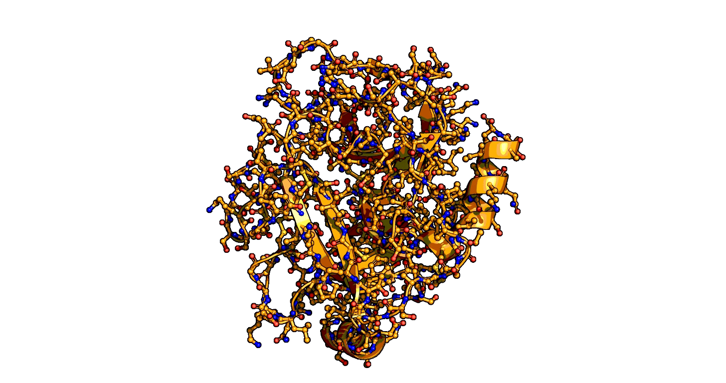
Using a series of more than 1,000 X-ray snapshots of the shapeshifting of enzymes in action, researchers at Stanford University have illuminated one of the great mysteries of life – how enzymes are able speed up life-sustaining biochemical reactions so dramatically. ...

“When I say enzymes speed up reactions, I mean as in a trillion-trillion times faster for some reactions,” ...

As a result, biochemists don’t have a basic understanding and, therefore, have been unable to predict enzyme rates or design new enzymes as well as nature does, an ability that would be impactful across industry and medicine.  ...

that enzymes are not a single structure. Instead, they focus on what they term “ensembles,” showing how enzymes move between different physical states – or conformational ensembles – during catalysis. ...

Exploring these ensembles and comparing reaction states on enzymes to states of uncatalyzed enzymes in pure water, ... broke down enzyme catalysis to the individual energetic contributions at the precise place where enzyme and target molecule meet during a reaction, known as the active site, to understand how they work chemically and physically to speed up reactions. ...

“Nature has evolved these mechanisms independently in multiple enzyme families – this is not an isolated feature, but catalytic mechanisms that have been discovered multiple times by nature through evolution. This means we may be able to copy nature and use this and other features to design and build new enzymes,” ..."

From the editor's summary and abstract:
"Editor’s summary
Three-dimensional structures of enzymes have provided important insights into the chemical mechanisms through which these catalysts achieve their incredible rate accelerations. Building on decades of enzymology and structural biology, Du et al. analyzed more than 1000 structures of serine proteases to develop a comprehensive and quantitative framework for the origins of rate acceleration in this enzyme family. The authors highlight precise positioning of catalytic residues, ground state destabilization, and motion that is flexible but restricted along a productive path as key factors in rate acceleration by serine proteases.  ...
Structured Abstract
INTRODUCTION
Enzymes are central to all biological functions. A century of enzymology has established chemical mechanisms and roles of active-site residues and cofactors for almost all known enzymes.
Yet, we still lack the ability to account for the enormous rate enhancements provided by enzymes.
Although serine proteases are often used as examples in biochemistry textbooks to illustrate enzyme catalysis, it remains unclear how their active sites accelerate the peptide bond cleavage reaction.
The serine nucleophile is a hydroxyl group that is similar to water in reactivity; the oxyanion hole typically consists of amide groups, which are also similar to water as hydrogen bond donors. The general base in the catalytic triad does facilitate proton transfer but not nearly enough to account for serine protease catalysis. A deepened understanding of how such enzymes work requires an approach that can identify catalytic features, uncover the physical basis of their function, and quantify their catalytic contributions.
RATIONALE
Reaction rates—and catalysis—are determined by the probability of achieving the reaction’s transition state, and these probabilities are defined by free energies. Conformational ensembles encode states and their probabilities, thereby providing a means to access the free energies that define catalysis.
We therefore moved beyond traditional structure-function relationships to develop an ensemble-function approach that identifies catalytic features and quantifies the catalytic contribution from each individual feature.
We applied this approach to serine proteases to address the following questions:
(i) What are the molecular changes between the enzymatic reaction states?
(ii) How does the enzymatic reaction differ from that in solution? and
(iii) What are the energetic consequences of these differences?
RESULTS
We brought together >1000 x-ray crystallographic structures from 17 serine proteases to build pseudo-ensembles, and we obtained ensemble information for the uncatalyzed solution reaction from small-molecule structures and computation. Pseudo-ensembles for enzymes bound to ground state analogs, transition state analogs, or no substrates revealed an altered energy landscape for the enzymatic reaction, where shorter and more efficient reaction paths are favored relative to those in solution.
The enzymes position reactants within a short distance and use a side-chain rotation of the catalytic serine to facilitate reaction, removing much of the translational motion needed in solution and increasing the reaction probability. The enzymatic reaction is further accelerated by multiple interactions that are destabilized in the ground state and relieved in the transition state.
We quantified catalytic contributions from these features using knowledge-based energy functions to provide a minimal model that accounts for serine protease catalysis within error. The same catalytic features are commonly used by enzymes from distinct structural folds to catalyze reactions that involve nucleophilic addition on carbonyl compounds, suggesting convergent evolution under the same mechanistic constraints and high evolvability of the identified strategies.
CONCLUSION
Ensemble-function analyses identified precise catalytic features and established an experimentally based, quantitative model for serine protease catalysis. This model is grounded in the most basic concepts of chemistry and physics—torsion angles, van der Waals interactions, hydrogen bonds, and conformational entropy—and thus is accessible to researchers and students alike. The ensemble-function approach connects these fundamental physicochemical properties to the emergent functions of macromolecules, a connection that is needed to understand enzyme catalysis and still more complex protein functions, such as allostery and molecular machines."

New findings on the power of enzymes could reshape biochemistry | Stanford Report


Animation showing the transition state of a serine protease – an example of how enzymes are always in motion.


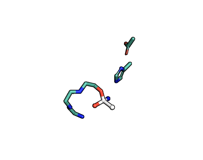
A close-up showing the serine protease reaction at the active site, place where enzyme and target molecule meet during a reaction.


The ensemble-function approach provides a quantitative catalytic model for serine proteases and identifies repeatedly evolved catalytic strategies.


Sunday, June 16, 2024

How our 2-billion-year-old microbial ancestors archaea made energy with hydrogen

Amazing stuff!

Will hydrogen become a viable alternative source of energy?

With hydrogen keep in mind: The Hindenburg Disaster of 1937! Hydrogen is dangerous, but human ingenuity will eventually learn to handle hydrogen safely.

"New research has redefined our understanding of how archaea – our 2-billion-year-old microbial ancestors – used hydrogen gas to produce energy. ...
There are 3 main domains of life on Earth: Eukarya, Archaea and Bacteria. ...
The team analysed the genomes of thousands of archaea and found they use unusual enzymes called [FeFe]-hydrogenases. Before this research, it was thought these enzymes were only produced by eukaryotes and bacteria.
And the archaeans’ hydrogen-using enzymes are the smallest and most complex. ..."

"... The findings, published today in Cell, explain how these tiny lifeforms make energy by consuming and producing hydrogen. This simple but dependable strategy has allowed them to thrive in some of Earth’s most hostile environments for billions of years. ...
The most widely accepted scientific theory also suggests that eukaryotes, such as humans, evolved from a very ancient lineage of archaea merging with a bacteria cell through exchanging hydrogen gas. ...
The team analysed the genomes of thousands of archaea for hydrogen-producing enzymes and then produced the enzymes in the lab to study their characteristics. They discovered that some archaea use unusual types of enzymes called [FeFe]-hydrogenases.

The archaea making these hydrogen-using enzymes were found in many of Earth’s most challenging environments, including hot springs, oil reservoirs, and deep beneath the seafloor. ..."

From the highlights and abstract:
"Highlights
Archaea from nine different phyla encode structurally diverse [FeFe] hydrogenases
• Active ultraminimal [FeFe] hydrogenases are produced by uncultured DPANN archaea
Ancient hybrid [FeFe] and [NiFe] hydrogenases are encoded by diverse archaea
Hydrogen-producing Asgard archaeal cultures express [FeFe] hydrogenases
Summary
Microbial hydrogen (H2) cycling underpins the diversity and functionality of diverse anoxic ecosystems. Among the three evolutionarily distinct hydrogenase superfamilies responsible, [FeFe] hydrogenases were thought to be restricted to bacteria and eukaryotes. Here, we show that anaerobic archaea encode diverse, active, and ancient lineages of [FeFe] hydrogenases through combining analysis of existing and new genomes with extensive biochemical experiments. [FeFe] hydrogenases are encoded by genomes of nine archaeal phyla and expressed by H2-producing Asgard archaeon cultures. We report an ultraminimal hydrogenase in DPANN archaea that binds the catalytic H-cluster and produces H2. Moreover, we identify and characterize remarkable hybrid complexes formed through the fusion of [FeFe] and [NiFe] hydrogenases in ten other archaeal orders. Phylogenetic analysis and structural modeling suggest a deep evolutionary history of hybrid hydrogenases. These findings reveal new metabolic adaptations of archaea, streamlined H2 catalysts for biotechnological development, and a surprisingly intertwined evolutionary history between the two major H2-metabolizing enzymes."

How our 2-billion-year-old microbial ancestors archaea made energy with hydrogen


Graphical abstract


Figure 1 Phylogenetically and metabolically diverse archaea encode [FeFe] hydrogenases


Sunday, February 11, 2024

An enzyme used in laundry detergent can recycle single-use bioplastics within 24 hours

Human ingenuity can handle plastic waste and recycling! No need for alarmism and hysteria over plastic!

From the highlights and abstract:
"Highlights
• Chemical modification of lipase enhances stability and activity in ionic liquids
• Ionic liquids allow for rapid chemoenzymatic depolymerization of PLA at 90°C
Post-consumer PLA fully water soluble within 24 h
• Post-consumer PLA fully converted to monomer within 40 h
Summary
The accumulation of plastic waste in the environment is an ecological disaster that requires a plurality of approaches to tackle. There is therefore an ever-pressing need to close the loop on production of both conventional and bioderived plastics. In response, we propose a generalizable biocatalysis engineering strategy to enhance the use of enzymes to depolymerize a broad class of plastics. To demonstrate this approach, we have solubilized and stabilized the commonly available lipase B from Candida antarctica in ionic liquids. In doing so, we unlock the ability of the enzyme to hydrolytically depolymerize post-consumer poly(lactic acid) plastic, demonstrating full degradation within 24 h and full conversion to monomer within 40 h at 90°C. This facile and scalable modification strategy allows for elevated operation temperatures in combination with the superior solvent capabilities of ionic liquids, providing a blueprint for enhancing the capability of any hydrolytic enzyme for plastic recycling."

An enzyme used in laundry detergent can recycle single-use plastics within 24 hours Scientists at King's College London have developed an innovative solution for recycling single-use bioplastics commonly used in disposable items such as coffee cups and food containers.


Graphical abstract


Monday, February 13, 2023

Building Better Enzymes through combinatorial assembly from modular building blocks

Good news!

"... a computational method for designing thousands of different active enzymes with unprecedented efficiency by assembling them from engineered modular building blocks. ...
The inspiration for this new approach came from within: our immune system, which is capable of making billions of different antibodies – proteins that in principle can counter any harmful microorganism – just from the bits dictated by a relatively small number of genes. “Antibodies are the only family of proteins in nature known to be generated in a modular way,” ... “Their huge diversity is achieved by recombining preexisting genetic fragments ..."

"Fishing for the right puzzle piece
Recombination can be a good strategy to generate natural protein diversity while retaining function, but it also causes problems if the starting sequences are too dissimilar and cannot fit together properly to form a functional protein. [researchers]  developed a machine learning strategy to piece together fragments sourced from highly divergent natural enzymes to generate a million structurally diverse protein backbones. This step is then followed by mutagenesis and structural optimization to create stable, functional active sites. Isolation by high-throughput yeast display and activity-based profiling recovered thousands of functional enzyme variants. A second-generation model trained on preorganization of the active site was nearly 10-fold more efficient and provides valuable insights for enzyme design strategies across the board."

From the abstract:
"The design of structurally diverse enzymes is constrained by long-range interactions that are necessary for accurate folding. We introduce an atomistic and machine learning strategy for the combinatorial assembly and design of enzymes (CADENZ) to design fragments that combine with one another to generate diverse, low-energy structures with stable catalytic constellations. We applied CADENZ to endoxylanases and used activity-based protein profiling to recover thousands of structurally diverse enzymes. Functional designs exhibit high active-site preorganization and more stable and compact packing outside the active site. Implementing these lessons into CADENZ led to a 10-fold improved hit rate and more than 10,000 recovered enzymes. This design-test-learn loop can be applied, in principle, to any modular protein family, yielding huge diversity and general lessons on protein design principles."

Building Better Enzymes – by Breaking Them Down - Chemistry | Weizmann Wonder Wander - News, Features and Discoveries In a major step toward greener industry, Weizmann Institute scientists create a computational method for generating enzymes with unprecedented efficiency


Here is the link to the preprint: https://www.biorxiv.org/node/2896090.full

Fig. 1. Key steps in the CADENZ workflow.


Friday, November 25, 2022

Researchers created artificial enzymes programmed to target the genetic code of SARS-CoV-2 or cancer and destroy the virus or cancer

Amazing stuff! Sounds very promising!

"... Although most enzymes are proteins, some of these crucial reactions are catalysed by RNA, a chemical cousin of DNA, which can fold into enzymes known as ribozymes. Some classes of ribozyme are able to target specific sequences in other RNA molecules and cut them precisely.
In 2014, ... discovered that artificial genetic material known as XNA – in other words, synthetic chemical alternatives to RNA and DNA not found in nature – could be used to create the world’s first fully-artificial enzymes ... named XNAzymes.
At the beginning, XNAzymes were inefficient, requiring unrealistic laboratory conditions to function. Earlier this year, however, ... a new generation of XNAzymes, engineered to be much more stable and efficient under conditions inside cells. These artificial enzymes can cut long, complex RNA molecules and are so precise that if the target sequence differs by just a single nucleotide (the basic structural unit of RNA), they will recognise not to cut it. This means they can be programmed to attack mutated RNAs involved in cancer or other diseases, leaving normal RNA molecules well alone. ...
Now, in research ... report how they have used this technology to successfully ‘kill’ live SARS-CoV-2 virus. ... 
“Put simply, XNAzymes are molecular scissors which recognise a particular sequence in the RNA, then chop it up. As soon as scientists published the RNA sequence of SARS-CoV-2, we started scanning through looking for sequences for our XNAzymes to attack.”
While these artificial enzymes can be programmed to recognise specific RNA sequences, the catalytic core of the XNAzyme – the machinery that operates the ‘scissors’ – does not change. This means that creating new XNAzymes can be done in far less time than it normally takes to develop antiviral drugs. ...
“It’s worth remembering, however, that the amazingly successful Pfizer and Moderna COVID-19 vaccines are themselves based on synthetic RNA molecules – so it’s a really exciting and rapidly developing field, with enormous potential.” ..."

From the abstract:
"The unprecedented emergence and spread of SARS-CoV-2, the coronavirus responsible for the COVID-19 pandemic, underscores the need for diagnostic and therapeutic technologies that can be rapidly tailored to novel threats. Here, we show that site-specific RNA endonuclease XNAzymes – artificial catalysts composed of single-stranded synthetic xeno-nucleic acid oligonucleotides (in this case 2’-deoxy-2’-fluoro-β-D-arabino nucleic acid) – may be designed, synthesised and screened within days, enabling the discovery of a range of enzymes targeting SARS-CoV-2 ORF1ab, ORF7b, spike- and nucleocapsid-encoding RNA. Three of these are further engineered to self-assemble into a catalytic nanostructure with enhanced biostability. This XNA nanostructure is capable of cleaving genomic SARS-CoV-2 RNA under physiological conditions, and when transfected into cells inhibits infection with authentic SARS-CoV-2 virus by RNA knockdown. These results demonstrate the potential of XNAzymes to provide a platform for the rapid generation of antiviral reagents."

‘Programmable molecular scissors’ could help fight COVID-19 infection | University of Cambridge Cambridge scientists have used synthetic biology to create artificial enzymes programmed to target the genetic code of SARS-CoV-2 and destroy the virus, an approach that could be used to develop a new generation of antiviral drugs.


Fig. 1: RNA endonuclease XNAzymes retargeted to the SARS-CoV-2 genome.


Saturday, November 05, 2022

Nanosensors target enzymes to monitor and study cancer

Good news! Cancer is history (soon)! Treating cancer at nanoscale precision with multiscale (space and time) analysis! Wow! Very impressive work! This is only the beginning!

It appears humans are finally understanding and outsmarting cancer at nanoscale!!!

"... Sensitive tools for measuring protein or gene expression, even on the single cell level, have helped researchers understand the different cell types present in a tumor’s microenvironment and how this composition changes after treatments. However, these assays don’t necessarily show which proteins are active or relevant to tumor progression, or allow clinicians to noninvasively monitor the progress of the disease or its response to treatment. A protein could be present in a cancer cell as a bystander, for example, but not an active participant in its cellular transformations. Enzymes, which catalyze biochemical reactions inside cells, may give a clearer picture of which genes or proteins to target at a particular time. ...
researchers ... have developed a set of enzyme-targeting nanoscale tools to monitor cancer progression and treatment response in real time, map enzyme activity to precise locations within a tumor, and isolate relevant cell populations for analysis. ...
the nanosensors could be used by clinicians to tailor treatments to a patient’s specific cancer, and to monitor cancer progression and treatment response, while researchers could use them to better understand the molecular biology of cancer and develop new tools to diagnose, track, and treat the disease ...
For several years, the ... laboratory has been developing noninvasive urine tests for the detection of cancer, including colon, ovarian, and lung cancer. The tests rely on nanoparticles that interact with tumor proteins called proteases. Proteases are a type of enzyme that act as molecular scissors to cleave proteins and break them down into smaller components. Proteases help cancer cells escape from tumors by cutting through the extracellular network of proteins that holds cells in place.

The nanoparticles are coated with peptides (short protein fragments) that target cancer-linked proteases. When the nanoparticles arrive at the tumor site, the peptides are cut and release biomarkers that can be detected in the urine.

In the current study, the researchers tested whether they could use this technology not just to detect cancer, but to track the development of cancer and its response to treatments accurately and sensitively over time. The team created a panel of 14 nanoparticles designed to target proteases overexpressed in non-small cell lung cancer induced in a mouse model. These nanoparticles had been adapted to release barcoded peptides when they encounter dysregulated enzymes in the tumor microenvironment.
Each nanosensor was able to track different patterns of protease activity, which changed dramatically as the tumor progressed. After treatment with a lung cancer-targeting drug, the researchers were able to find signs tumor regression quickly, within just three days of administering treatment. ...
Having identified nanosensors of interest, researchers mapped where in the tumor microenvironment the enzymes acting on these sensors were active. They adapted their nanoprobes to leave behind fluorescent tags when they are cleaved from the nanosensor, assigning different tags to different proteases. After applying the nanoprobes to samples of lung tissue, they looked for patterns in how the tags were distributed.

One tag resulted in a curious spindle-like pattern that turned out to belong to the tumor vasculature. Researchers pinpointed the protease activity to specific types of cells: endothelial cells, which line blood vessels, and pericytes, which regulate vascular function and are actively recruited in angiogenesis — one of the archetypal hallmarks of cancer cell growth. ...
Ultimately, however, the team envisions panels of nanoprobes targeting several important features of cancer simultaneously and noninvasively in patients. Other hallmarks of cancer include proliferative signaling, the evasion of growth suppressors, genome instability, resistance to cell death, deregulated metabolism, and activation of invasion and metastasis. Because cancer alters protease activity across all of these processes, the team’s nanoprobes could be designed to target these different processes, with the aim of providing a comprehensive picture of tumor activity driving the disease. ..."

From the abstract:
"Diverse processes in cancer are mediated by enzymes, which most proximally exert their function through their activity. High-fidelity methods to profile enzyme activity are therefore critical to understanding and targeting the pathological roles of enzymes in cancer. Here, we present an integrated set of methods for measuring specific protease activities across scales, and deploy these methods to study treatment response in an autochthonous model of Alk-mutant lung cancer. We leverage multiplexed nanosensors and machine learning to analyze in vivo protease activity dynamics in lung cancer, identifying significant dysregulation that includes enhanced cleavage of a peptide, S1, which rapidly returns to healthy levels with targeted therapy. Through direct on-tissue localization of protease activity, we pinpoint S1 cleavage to the tumor vasculature. To link protease activity to cellular function, we design a high-throughput method to isolate and characterize proteolytically active cells, uncovering a pro-angiogenic phenotype in S1-cleaving cells. These methods provide a framework for functional, multiscale characterization of protease dysregulation in cancer."

Nanosensors target enzymes to monitor and study cancer | MIT News | Massachusetts Institute of Technology By analyzing enzyme activity at the organism, tissue, and cellular scales, new sensors could provide new tools to clinicians and cancer researchers.


Fig. 1: Multiscale profiling of protease activity in cancer


Saturday, September 03, 2022

Study finds enzyme in the brain is a regulator for body weight

Good news!

"... In a new study, they found that removing the enzyme from neurons in a part of the brain known as the hypothalamus led mice to gain weight and burn less fat. This finding, they say, suggests that the enzyme could be a target for treating metabolic disease. ...
For the study, Yale researchers focused on an enzyme called O-linked b-D-N-acetylglucosamine transferase, or OGT. ...
As a first step, researchers observed what happened to OGT in neurons of the ventromedial hypothalamus when food intake was adjusted. They found that when mice consumed less food, OGT levels went up. ...
... bred mice that lacked OGT in neurons of the ventromedial hypothalamus. They found that the mice gained weight very quickly on a normal diet, becoming much heavier than typical mice even though they were eating the same amount of food and were just as physically active. ...
But in the study, mice lacking OGT didn’t burn fat as much as other mice when food was restricted. ..."

From the abstract:
"The ventromedial hypothalamus (VMH) is known to regulate body weight and counterregulatory response. However, how VMH neurons regulate lipid metabolism and energy balance remains unknown. O-linked β-D-N-acetylglucosamine (O-GlcNAc) modification (O-GlcNAcylation), catalyzed by O-GlcNAc transferase (OGT), is considered a cellular sensor of nutrients and hormones. Here, we report that genetic ablation of OGT in VMH neurons inhibits neuronal excitability. Mice with VMH neuron-specific OGT deletion show rapid weight gain, increased adiposity, and reduced energy expenditure, without significant changes in food intake or physical activity. The obesity phenotype is associated with adipocyte hypertrophy and reduced lipolysis of white adipose tissues. In addition, OGT deletion in VMH neurons down-regulates the sympathetic activity and impairs the sympathetic innervation of white adipose tissues. These findings identify OGT in the VMH as a homeostatic set point that controls body weight and underscore the importance of the VMH in regulating lipid metabolism through white adipose tissue–specific innervation."

Study finds enzyme in the brain is a ‘metastat’ for body weight | YaleNews


Fig. 8. OGT is required for the neuronal activity of the VMH.


Tuesday, August 23, 2022

One Enzyme Likely Responsible for Mutations in Most Cancers

Good news! Cancer is history (soon)!

"Over half of all the cancer genomes that researchers have sequenced share one characteristic: They contain mutational signatures associated with a family of enzymes called APOBEC3, suggesting a role for these enzymes in mutagenesis. So far, limited evidence suggests that the main culprit within the APOBEC3 family is the enzyme APOBEC3B. Though much about the enzyme’s activity remains unknown, the field largely focused on it as the primary enzyme responsible for cancer mutations. But APOBEC3A—previously thought not to play such a prominent role—may actually be responsible for the mutations seen in most cancer cell lines, a paper published July 20 in Nature suggests, making it a potential therapeutic target in cancer. ...
APOBEC3 enzymes’ main role is to induce mutations in viral RNA and DNA that stop pathogenic viruses from replicating. However, APOBEC3 enzymes have also been implicated in cancer, as some of the patterns of mutations discovered in human cancer genomes, called ‘mutational signatures’, were found to resemble mutational patterns that some APOBEC3 enzymes induce on viral nucleic acids. And this applies to more than just a few cancers: The mutational signatures associated with APOBEC activities, in particular that of the APOBEC3 subfamily, have been found “in more than 50 percent of all cancer genomes looked at up to date and more than 70 percent of cancer types,” ..."

From the abstract:
"The APOBEC3 family of cytosine deaminases has been implicated in some of the most prevalent mutational signatures in cancer. However, a causal link between endogenous APOBEC3 enzymes and mutational signatures in human cancer genomes has not been established, leaving the mechanisms of APOBEC3 mutagenesis poorly understood. Here, to investigate the mechanisms of APOBEC3 mutagenesis, we deleted implicated genes from human cancer cell lines that naturally generate APOBEC3-associated mutational signatures over time. Analysis of non-clustered and clustered signatures across whole-genome sequences from 251 breast, bladder and lymphoma cancer cell line clones revealed that APOBEC3A deletion diminished APOBEC3-associated mutational signatures. Deletion of both APOBEC3A and APOBEC3B further decreased APOBEC3 mutation burdens, without eliminating them. Deletion of APOBEC3B increased APOBEC3A protein levels, activity and APOBEC3A-mediated mutagenesis in some cell lines. The uracil glycosylase UNG was required for APOBEC3-mediated transversions, whereas the loss of the translesion polymerase REV1 decreased overall mutation burdens. Together, these data represent direct evidence that endogenous APOBEC3 deaminases generate prevalent mutational signatures in human cancer cells. Our results identify APOBEC3A as the main driver of these mutations, indicate that APOBEC3B can restrain APOBEC3A-dependent mutagenesis while contributing its own smaller mutation burdens and dissect mechanisms that translate APOBEC3 activities into distinct mutational signatures."

Underdog Enzyme Likely Responsible for Mutations in Most Cancers | The Scientist Magazine® A previously overlooked enzyme called APOBEC3A is linked to the most prevalent mutational signatures in cancer cell lines, a study finds.


Fig. 4: APOBEC3 deaminases drive the acquisition of clustered mutations in human cancer cells


Wednesday, August 03, 2022

Anaerobic bacteria can convert hydrogen and CO2 to formic acid

Contra the Global Warming Hoax and Climate Change Religion propaganda and demagoguery! Human ingenuity can easily handle any carbon dioxide in the atmosphere!

Apparently, these anaerobic bacteria can convert hydrogen and CO2 to formic acid. This solution would solve two issues at the same time: hydrogen storage and CO2 removal.

One of the authors of the research article below has actually authored a series of articles on the same bacteria and its amazing capabilities.

From the abstract:
"Filamentous enzymes have been found in all domains of life, but the advantage of filamentation is often elusive. Some anaerobic, autotrophic bacteria have an unusual filamentous enzyme for CO2 fixationhydrogen-dependent CO2 reductase (HDCR)2,3—which directly converts H2 and CO2 into formic acid. HDCR reduces CO2 with a higher activity than any other known biological or chemical catalyst, and it has therefore gained considerable interest in two areas of global relevance: hydrogen storage and combating climate change by capturing atmospheric CO2. However, the mechanistic basis of the high catalytic turnover rate of HDCR has remained unknown. Here we use cryo-electron microscopy to reveal the structure of a short HDCR filament from the acetogenic bacterium Thermoanaerobacter kivui. The minimum repeating unit is a hexamer that consists of a formate dehydrogenase (FdhF) and two hydrogenases (HydA2) bound around a central core of hydrogenase Fe-S subunits, one HycB3 and two HycB4. These small bacterial polyferredoxin-like proteins oligomerize through their C-terminal helices to form the backbone of the filament. By combining structure-directed mutagenesis with enzymatic analysis, we show that filamentation and rapid electron transfer through the filament enhance the activity of HDCR. To investigate the structure of HDCR in situ, we imaged T. kivui cells with cryo-electron tomography and found that HDCR filaments bundle into large ring-shaped superstructures attached to the plasma membrane. This supramolecular organization may further enhance the stability and connectivity of HDCR to form a specialized metabolic subcompartment within the cell."

Bakterien wandeln Kohlendioxid und Wasserstoff in Ameisensäure um Wärmeliebende Bakterien nutzen CO₂ und molekularen Wasserstoff als Energiequelle – und bilden daraus Ameisensäure. Forschern ist es nun gelungen, die molekularen Details dieses biotechnologisch aussichtsreichen Prozesses zu entschlüsseln.

Membrane-anchored HDCR nanowires drive hydrogen-powered CO2 fixation (no public access, but article above contains link to PDF file)

Saturday, July 20, 2019

Without LPCAT1 Multiple Types Of Tumors Cannot Survive

Posted: 7/20/2019

Are we finally closing in to defeat cancer! Yes we are! Here is more evidence! Cancer is history!

“[DNA sequencing] Complementary biochemical and biophysical perspectives of cancer point toward profound shifts in nutrient uptake and utilization that propel tumor growth and major changes in the structure of the plasma membrane of tumor cells.” (S3; emphasis added)
The more targets, the better the offensive! 

We should have very powerful cocktails or other therapies (e.g. immunotherapies) in 10-20 years that will make the eradication of cancer possible. We may also opt to change some genetic code to prevent cancer! Not to mention that our diagnostic tools are getting so much better like liquid biopsy of blood etc.

Sources (S):