Showing posts with label ophthalmology. Show all posts

Monday, July 27, 2026

European and US regulators have approved a retinal implant and camera equipped glasses to improve irreversible blindness

Good news!

When can I get my eagle eyes and fine print reader? 😊

As an aside, in German prima means great!

"... retinal implant, which combines a tiny chip inserted beneath the retina with camera-equipped glasses to restore functional vision"

"“Science Corporation, a startup developing novel brain-computer interfaces (BCI), won approval from Europe’s medical device regulator to begin selling a device that restores vision lost from age-related macular degeneration.

The company said the device, called PRIMA, also received a designation from the US Food and Drug Administration that is the first step toward an expedited regulatory review, which could see the device used to treat two rare kinds of blindness.”"

From the abstract:
"Background
Geographic atrophy (GA) due to age-related macular degeneration (AMD) is the leading cause of irreversible blindness and affects over 5 million people globally. Currently, no therapies exist to restore vision to the affected persons. The PRIMA system combines a subretinal photovoltaic implant and near-infrared light-projecting glasses to restore sight to areas of central retinal atrophy.

Methods
In an open-label, baseline-controlled, multicenter, prospective, single-arm clinical study, the vision of participants with GA and a visual acuity of logMAR ≥1.2 was assessed with and without PRIMA glasses at six and twelve months.
The primary endpoints were the proportion of participants with improvement in visual acuity (VA) of logMAR 0.2 or more and the number and severity of study-related serious adverse events at 12 months.

Results
Thirty-two of thirty-eight enrolled participants reached the 12-month endpoint. Of the six who did not, three died, one withdrew, and two were unavailable for testing. Twenty-six (81.3%; 95% CI: 63.56–92.79%) had an improved prosthetic VA of at least logMAR 0.2 versus baseline (p<0.001) and
in 19 participants, there were 26 serious adverse events. Twenty-one of these events (81%) occurred within the first two months, of which 20 (95%) were resolved within two months of implantation. There was no significant change in peripheral natural VA compared to baseline.

Conclusions
In this study of a limited number of participants, the PRIMA system restored central vision with significant improvements of VA in participants with GA due to AMD. ..."

Doomslayer: Progress Roundup - by Malcolm Cochran


The glasses
The implant


Figure 1. Rendering of the PRIMA system components.


Sunday, July 12, 2026

First 3D Structure of Inactivated Cone Opsins Revealed in three different studies

Amazing stuff! I limit myself here to one of the three papers.

"... In a new study, ... two researchers ... have succeeded for the first time in determining the three-dimensional structure of human cone opsins in their dark state and showing how their molecular architecture enables their rapid activation by light. This provides important new insights into human vision and its evolution and may offer new starting points for the study of eye diseases that currently lack effective treatment. ..."

From the editor's summary and abstract:
"Editor’s summary
Human daytime vision relies on a trio of visual receptors called opsins, which are found in the cone cells in and around the central region of the retina.
The three opsins are tuned to long, medium, or short wavelengths of light, roughly corresponding to red, green, and blue, and mutations or other defects in cone cell function can lead to vision deficits.
Although the cell biology and biochemistry of color vision have been well studied, up to now, the molecular explanation for cone opsin spectral tuning and signaling kinetics has been limited by a lack of experimental structures.
Three papers in this issue now resolve this deficit.
Schmidt et al. determined structures of the dark state of the green and blue human cone opsins, which revealed important details of these receptors and provide a basis for a femtosecond-resolution spectroscopy study.
Ohashi et al. performed complementary structural, spectroscopic, and computational results with dark-state red and green cone opsins from macaques, which have color vision similar to humans.
Finally, Peng et al. studied all three human cone opsins in the presumed active state bound to a G protein and all-trans retinal.
The three papers together provide a clear picture of the features of these visual receptors that lead to different spectral properties, activation and inactivation kinetics, and recycling. ...

Structured Abstract
INTRODUCTION
High-acuity daylight vision relies on cone photoreceptors, specialized class A G protein–coupled receptors (GPCRs). Like other light-sensitive GPCRs, the three human cone opsins covalently bind vitamin A derivative 11-cis-retinal through a protonated Schiff base. Despite sharing the same chromophore, they detect distinct wavelengths of light and generate swift signaling responses at high repetition rates. Although cone opsins are central to human vision, and in contrast to the well-studied rod photoreceptor rhodopsin, the detailed molecular basis of these functional specializations remains elusive.

RATIONALE
To obtain structure-function relationships that extend the kinetic and mechanistic understanding of photopic vision, we solved cryo–electron microscopy (cryo-EM) structures of the two most evolutionarily and functionally divergent human cone opsins, short-wavelength-sensitive OPN1SW and medium-wavelength-sensitive OPN1MW, in their initial 11-cis-coupled state. We combined the structural data with multiple functional assays, hybrid quantum mechanics/molecular mechanics simulations, time-resolved spectroscopy, and multitaxon opsin sequence analysis.

RESULTS
Cryo-EM structures of cone opsins revealed receptor-specific activation mechanisms and distinct strategies for stabilizing the retinal Schiff base.
OPN1SW, representing the phylogenetically older vertebrate opsins, has a more constrained polar chromophore environment, which contributes to its blue-shifted maximum absorption wavelength (λmax), yet its stabilization is weaker than that of rhodopsin. The architecture of OPN1SW shows substantial divergences in the canonical GPCR microswitch networks, including the replacement of the toggle switch with Y6.48, a disrupted PIF triad, and the absence of a highly conserved sodium- or water-coordination site.
Collectively, these alterations favor a preactive conformation, also captured by cryo-EM. OPN1SW further uses W185ECL2 as a steric switch to transmit the retinal isomerization event across several helices through an extended aromatic network.
In contrast, OPN1MW contains a chloride ion within the chromophore-binding pocket that modulates wavelength sensitivity and influences the amplitude of G protein signaling. This chloride-binding site coevolved with a structural pathway on helix 2 that couples chromophore chemistry to canonical GPCR microswitches.
Both receptors have accessible binding pockets that allow rapid ligand hydrolysis and, consequently, fast retinal turnover.
Femtosecond transient-absorption spectroscopy resolved the photoisomerization cascade, supporting a model in which deprotonation and subsequent hydrolysis limit signal duration in cone opsins.

CONCLUSION
Our structural and mechanistic insights describe how distinctive chromophore environments and GPCR microswitch adaptations tune spectral sensitivity and signaling-state lifetimes in cone opsins. Conservation of central residues across short-wavelength-sensitive and medium-to-long-wavelength-sensitive opsins suggests shared mechanistic principles that shaped the evolution of daylight vision. Similar motifs in other GPCRs, including sensory receptors, inform the strategies for modulating receptor activation kinetics and signal duration."

First 3D Structure of Inactivated Cone Opsins Revealed | The Scientist "Scientists resolved the three-dimensional structures of light-sensitive cone opsins, offering new avenues for treating age-related vision loss."

New insights into human vision (original news release)

Experiment performed at ELI Enables New Insights into Human Vision (original news release) "The retina of the human eye contains six to seven million cone cells. These cells contain light-sensitive proteins known as cone opsins. They enable us to perceive our surroundings in detail in daylight. In a new study, researchers ... have now, for the first time, determined the three-dimensional molecular structure of human cone opsins in their dark state, that is, before they are activated by light."


Cone opsins use distinct chromophore-stabilization strategies to tune spectral sensitivity, activation kinetics, and retinal regeneration.


Sunday, July 05, 2026

Lab-grown retinal cells show promise for new eye therapies

Good news! This could be a breakthrough! Make the blind see again!

"Biomedical engineers ... have used induced pluripotent stem cells (iPSCs) to grow specialized blood vessel cells critical to retinal health for the first time. When injected into mouse models of retinal disease, these "retinal endothelial cells" integrated into the damaged tissue to regenerate blood vessels and restore retinal function. Researchers also demonstrated the cells' ability to form functional retinal vascular tissue in a lab-grown environment, providing a pathway to model and research various eye diseases. ..."

From the abstract:
"Retinal microvascular diseases involve a compromised inner blood–retina barrier (iBRB), which remains poorly understood. A renewable source of human iBRB endothelium is thus vital for advancing eye research and treatment development.
Here we differentiated human induced pluripotent stem cells into retinal endothelial cells (iRECs) via the Wnt–β-catenin pathway, namely Norrin–Frizzled4 signalling.
These iRECs show genetic, protein and functional fidelity as well as unique retinal features.
When injected into oxygen-induced retinopathy mice, iRECs integrated into the host vascular network and revascularized the ischaemic eye, rescuing the tissue.
In microphysiological models, iRECs form perfusable microvascular networks that recapitulate iBRB morphology and phenotype in both healthy and diabetic states while also physiologically organizing and interacting with induced pluripotent stem cell-derived retinal pericytes.
Our study establishes functional human iRECs and microphysiological iBRB models that facilitate mechanistic studies aimed at identifying therapeutic targets and promoting the revascularization of injured retinas, thereby supporting treatment advancement."

Lab-grown retinal cells show promise for new eye therapies

Lab-Grown Retinal Cells Show Promise for New Eye Therapies (original news release) "Critical cells that line retinal blood vessels grown from stem cells restore retinal function in mouse models and form retinal tissue in a lab for future disease studies."


This image depicts a mouse’s retina suffering from conditions similar to diabetic retinopathy both before (right) and after (left) being treated with human lab-grown retinal endothelial cells.
The green in the left image shows the human lab-grown retinal endothelial cells integrating into the damaged mouse retina, demonstrating their potential use to treat early stages of the disease.


Fig. 1: Derivation of retinal endothelial cells from human induced pluripotent stem cells via the Norrin–Fz4 axis.


Fig. 4: iRECs revascularize the ischaemic eye.


Wednesday, December 03, 2025

3D-printed cornea restores blind patient's sight for the first time

Good news!

"In a major breakthrough in human tissue replication, for the first time ever a 3D-printed cornea has been transplanted onto a legally blind patient's eye, successfully restoring their sight. ..."

"In late October 2025, Rambam Eye Institute's Cornea Unit performed the world’s first transplantation of a fully 3D-bio-fabricated, cell-based corneal implant. The procedure was carried out on a patient who was legally blind in the treated eye, marking the first time anywhere that a corneal implant grown entirely from cultured human corneal cells, rather than donor tissue, has been successfully transplanted in a human being. In this instance, a single cornea from a healthy, deceased donor was cultured in the lab to create and print an additional 300 corneal implants. ..."

3D-printed cornea restores blind patient's sight for the first time


Monday, August 18, 2025

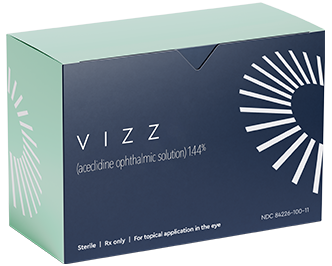
FDA approves VIZZ eye drops for presbyopia relief

Good news!

"The first aceclidine-based eye drop to improve near vision in adults with presbyopia, which affects more than 100 million adults in the US alone, has been approved by the Food and Drug Administration (FDA) and will be available within three months. ..."

FDA approves VIZZ eye drops for presbyopia relief


Saturday, March 29, 2025

Identification and characterization of human retinal stem cells capable of retinal regeneration

Good news!

"Human neural retinal stem-like cells (hNRSCs) found in fetal retinas and organoids show promise for retinal regeneration and vision restoration."

From the editor's summary and abstract:
"Editor’s summary
Cell-based therapies could potentially reverse retinal degeneration but require access to cells with high differentiation and safety profiles. Here, Liu et al. identified a distinct population of human neural retinal stem-like cells (hNRSCs) that reside in the ciliary marginal zone (CMZ) of fetal retinas and exhibit transcription profiles, indicating high differentiation and self-renewal potential. The authors found a similar cell type in a CMZ-like region of human retinal organoids (hROs). Transplanted hRO-derived hNRSCs differentiated into various retinal cell types and improved visual function in a mouse model of retinitis pigmentosa. hNRSCs could provide a useful and replenishable resource for translational research and therapeutic applications. ...

Abstract
Human retinal stem cells hold great promise in regenerative medicine, yet their existence and characteristics remain elusive.
Here, we performed single-cell multiomics and spatial transcriptomics of human fetal retinas and uncovered a cell subpopulation, human neural retinal stem-like cells (hNRSCs), distinct from retinal pigment epithelium stem-like cells and traditional retinal progenitor cells. We found that these hNRSCs reside in the peripheral retina in the ciliary marginal zone, exhibiting substantial self-renewal and differentiation potential.
We conducted single-cell and spatial transcriptomic analyses of human retinal organoids (hROs) and revealed that hROs contain a population of hNRSCs with similar transcriptional profiles and developmental trajectories to hNRSCs in the fetal retina potentially capable of regenerating all retinal cells.
Furthermore, we identified crucial transcription factors, such as MECOM, governing hNRSC commitment to neural retinogenesis and regulating repair processes in hROs. 
hRO-derived hNRSCs transplanted into the rd10 mouse model of retinitis pigmentosa differentiated and were integrated into the retina, alleviated retinal degeneration, and improved visual function.
Overall, our work identifies and characterizes a distinct category of retinal stem cells from human retinas, underscoring their regenerative potential and promise for transplantation therapy."

Contents | Science Translational Medicine 17, 791

Identification and characterization of human retinal stem cells capable of retinal regeneration (no public access) [This paper was apparently first published back in December 2023]

Monday, March 10, 2025

New hope for repairing eye damage once thought untreatable

Good news!

"A ... clinical trial of a procedure that took stem cells from a healthy eye and transplanted them into a damaged eye safely restored corneal surfaces in 14 patients who were followed for 18 months. 

The stem cell treatment for blinding cornea injuries — called cultivated autologous limbal epithelial cells, or CALEC ... It consists of removing stem cells from a healthy eye with a biopsy, expanding them into a cellular tissue graft in a novel manufacturing process that takes two to three weeks, and then surgically transplanting the graft into the eye with a damaged cornea. ...

One limitation of this approach is that it is necessary for the patient to have only one involved eye so a biopsy can be performed to get starting material from the unaffected normal eye. ...

The procedure remains experimental and is currently not offered ... at any U.S. hospital, and additional studies will be needed before the treatment is submitted for federal approval. ..."

New hope for repairing eye damage once thought untreatable — Harvard Gazette "Stem cell therapy safely restores cornea’s surface in clinical trial"

Sunday, October 27, 2024

Science Corporation restores eye sight of blind individuals with a brain-computer interface implant

Good news! Amazing stuff!

"Science Corporation, a company developing a brain-computer interface akin to Neuralink, recently announced the results of a clinical trial testing their implant's ability to restore vision to the blind. According to reporting from Wired, the trial participants started with an average visual acuity of 20/450, far below the legal blindness threshold of 20/200, and ended with an average of 20/160. In practical terms, the implant allowed a group of previously blind people to read, recognize faces, play cards, and solve crossword puzzles."

"... The [38] patients in the trials had lost their central visual field, which makes them unable to read and struggle to recognize faces. The results showed that the PRIMA implant restored real form vision in these patients such that sequences of letters can be read with a clinically meaningful improvement of letter acuity.

”The results demonstrate a milestone in the treatment of blindness caused by geographic atrophy due to age-related macular degeneration. For the first time it was possible to restore real form vision in a retina that has deteriorated due to age-related macular degeneration” ...

Patients show a notable improvement in their letter acuity while using the PRIMA implant to read a series of letters; some are able to read longer text. ...

The PRIMA system is a visual prosthesis which consists of a photovoltaic implant—totally cableless and autonomous—surgically implanted under the retina, a special pair of glasses with a camera and a projection system, and a pocket processor that processes the image for clarity and magnification. ..."

Weekly Progress Roundup - by Malcolm Cochran - Doomslayer


The PRIMA System Zoom is an assistive feature of the PRIMA system that changes the size of the features in the image on the retina and implant, but provides a correspondingly smaller field of view.


Thursday, September 26, 2024

More than 1 in 3 children around the world are shortsighted pointing to excessive screen time

Bad news! I am sure human ingenuity can handle the situation!

"More than 1 in 3 children around the world are shortsighted, per a large new study that points to excessive screen time and too little time outdoors; Asian children were most affected, with a prevalence rate of 85% in Japan and 73% in South Korea, versus 1% in Paraguay and Uganda. BBC"

From the abstract
"Background
Myopia is a pervasive global public health concern, particularly among the younger population. However, the escalating prevalence of myopia remains uncertain. Hence, our research aims to ascertain the global and regional prevalence of myopia, along with its occurrence within specific demographic groups.
Methods
An exhaustive literature search was performed on several databases covering the period from their inception to 27 June 2023. The global prevalence of myopia was determined by employing pooled estimates with a 95% CI, and further analysis was conducted to assess variations in prevalence estimates across different subgroups. Additionally, a time series model was utilised to forecast and fit accurately the future prevalence of myopia for the next three decades.
Results
This study encompasses a comprehensive analysis of 276 studies, involving a total of 5.410.945 participants from 50 countries across all six continents. The findings revealed a gradual increase in pooled prevalence of myopia, ranging from 24.32% (95% CI 15.23% to 33.40%) to 35.81% (95% CI 31.70% to 39.91%), observed from 1990 to 2023, and projections indicate that this prevalence is expected to reach 36.59% in 2040 and 39.80% in 2050. Notably, individuals residing in East Asia (35.22%) or in urban areas (28.55%), female gender (33.57%), adolescents (47.00%), and high school students (45.71%) exhibit a higher proportion of myopia prevalence.
Conclusion
The global prevalence of childhood myopia is substantial, affecting approximately one-third of children and adolescents, with notable variations in prevalence across different demographic groups. It is anticipated that the global incidence of myopia will exceed 740 million cases by 2050."

Global Health NOW: Revisiting Stockpiles as Mpox Spreads; The Power of the Promotoras Model; and DIY Injections

Thursday, May 16, 2024

Prevent myopia by staying more outdoors?

Is this the cause of my myopia? Staying indoors too much looking at a computer screen? (just kidding) 😊 But this has been suspected and reported for several decades!

Or why do bookworms wear glasses more frequently than others.

"Spending at least two hours outside is one of the best ways to prevent children from developing near-sightedness, or myopia, according to ophthalmologists. 
42% of people are myopic in the U.S.—up from 25% in the 1970s. 

Up to 90% are myopic in East Asia by young adulthood. 
Cause: Myopia occurs when the eyeball stretches, making faraway objects look blurry. But light stimulates the eye and releases dopamine, which can prevent stretching. 

Policy in practice: Pei-Chang Wu, a Taiwanese ophthalmologist, convinced Taiwan’s Ministry of Education to send elementary school students outdoors for at least two hours daily. 
Since the program’s implementation, myopia cases in primary school students have fallen for the first time in decades, from 50% in 2011 to 45.1% by 2015."

Global Health NOW: Heat’s ‘Far-Reaching’ Harm to Global Health; Helping Haitian Parents Help Kids; and Keeping Sight of Outdoor Time

Tuesday, April 16, 2024

AI makes retinal imaging 100 times faster, compared to manual method

Good news! Using a twin GAN!

"Researchers at the National Institutes of Health applied artificial intelligence (AI) to a technique that produces high-resolution images of cells in the eye. They report that with AI, imaging is 100 times faster and improves image contrast 3.5-fold. The advance, they say, will provide researchers with a better tool to evaluate age-related macular degeneration (AMD) and other retinal diseases. ...
By integrating AI with AO-OCT [adaptive optics, optical coherence tomography], ... that a major obstacle for routine clinical imaging using AO-OCT has been overcome, especially for diseases that affect the RPE [retinal pigment epithelium], which has traditionally been difficult to image. ..."

From the abstract:
"Background
In vivo imaging of the human retina using adaptive optics optical coherence tomography (AO-OCT) has transformed medical imaging by enabling visualization of 3D retinal structures at cellular-scale resolution, including the retinal pigment epithelial (RPE) cells, which are essential for maintaining visual function. However, because noise inherent to the imaging process (e.g., speckle) makes it difficult to visualize RPE cells from a single volume acquisition, a large number of 3D volumes are typically averaged to improve contrast, substantially increasing the acquisition duration and reducing the overall imaging throughput.
Methods
Here, we introduce parallel discriminator generative adversarial network (P-GAN), an artificial intelligence (AI) method designed to recover speckle-obscured cellular features from a single AO-OCT volume, circumventing the need for acquiring a large number of volumes for averaging. The combination of two parallel discriminators in P-GAN provides additional feedback to the generator to more faithfully recover both local and global cellular structures. Imaging data from 8 eyes of 7 participants were used in this study.
Results
We show that P-GAN not only improves RPE cell contrast by 3.5-fold, but also improves the end-to-end time required to visualize RPE cells by 99-fold, thereby enabling large-scale imaging of cells in the living human eye. RPE cell spacing measured across a large set of AI recovered images from 3 participants were in agreement with expected normative ranges.
Conclusions
The results demonstrate the potential of AI assisted imaging in overcoming a key limitation of RPE imaging and making it more accessible in a routine clinical setting."

AI makes retinal imaging 100 times faster, compared to manual method | National Institutes of Health (NIH) NIH scientists use artificial intelligence called ‘P-GAN’ to improve next-generation imaging of cells in the back of the eye.


Fig. 1: Overview of artificial intelligence (AI) enhanced retinal pigment epithelial (RPE) cell imaging strategy.

Fig. 4: Parallel discriminator generative adversarial network (P-GAN) enabled wide-scale visualization of the retinal pigment epithelial (RPE) cellular mosaic.


Sunday, February 18, 2024

Glasses with spiral lenses could help you see clearer, farther

Good news! Could be a gamechanger!

"... For the new study, scientists from the Photonics, Numerical and Nanosciences Laboratory (LP2N) in France have developed a new type of lens, which they call a “spiral diopter.” As the name suggests, this lens has a spiral shape, which creates three different focal points in the field of vision.

Unlike existing multifocal lenses, our lens performs well under a wide range of light conditions and maintains multifocality regardless of the size of the pupil,” ...  “For potential implant users or people with age-related farsightedness, it could provide consistently clear vision, potentially revolutionizing ophthalmology.” ..."

From the abstract:
"Lens design is of paramount importance in the evolving world of technology, where compactness and high optical performance are a necessity, ranging from smartphones and wearable devices to vehicles and virtual reality. Freeform design techniques allow us to transcend traditional limitations, but creating new optics remains a substantial challenge unless we consider unconventional physical phenomena. Here, we introduce a lens type based on freeform design, employing spiralization of one of its diopters that results in optical vortices. This enables multifocality, primarily serving needs in ophthalmology; however, its potential applications could broadly impact many other domains. In particular, this lens design could be crucial in miniaturizing emerging technologies while retaining their optical quality."

Glasses with spiral lenses could help you see clearer, farther Scientists have developed a new type of lens that creates multiple focal points, which could make for glasses or contacts that provide a clearer view over a range of distances. The secret? Making the lens a spiral shape.


Fig. 1. Comparison between (a) a conventional astigmatic lens and (b) our spiral lens. For each lens a ray tracing representation is given as well as a representation of the PSF in each expected focal zone.


Friday, January 12, 2024

Lab-grown retinas explain why people see colors dogs can't. Really!

What about animals that see more colors than humans?

What about all the scents a dog can smell and why humans can't? I want a dog nose! 😊

Or in other words, Johns Hopkins University has very poorly chosen a title for their article!

"... The findings ... increase understanding of color blindness, age-related vision loss, and other diseases linked to photoreceptor cells. They also demonstrate how genes instruct the human retina to make specific color-sensing cells, a process scientists thought was controlled by thyroid hormones.
By tweaking the cellular properties of the organoids, the research team found that a molecule derived from vitamin A called retinoic acid determines whether a cone will specialize in sensing red or green light. Only humans with normal vision and closely related primates develop the red sensor. ...
Instead, the new findings suggest red cones materialize through a specific sequence of events orchestrated by retinoic acid within the eye. ..."

From the abstract:
"Trichromacy is unique to primates among placental mammals, enabled by blue (short/S), green (medium/M), and red (long/L) cones. In humans, great apes, and Old World monkeys, cones make a poorly understood choice between M and L cone subtype fates. To determine mechanisms specifying M and L cones, we developed an approach to visualize expression of the highly similar M- and L-opsin mRNAs. M-opsin was observed before L-opsin expression during early human eye development, suggesting that M cones are generated before L cones. In adult human tissue, the early-developing central retina contained a mix of M and L cones compared to the late-developing peripheral region, which contained a high proportion of L cones. Retinoic acid (RA)-synthesizing enzymes are highly expressed early in retinal development. High RA signaling early was sufficient to promote M cone fate and suppress L cone fate in retinal organoids. Across a human population sample, natural variation in the ratios of M and L cone subtypes was associated with a noncoding polymorphism in the NR2F2 gene, a mediator of RA signaling. Our data suggest that RA promotes M cone fate early in development to generate the pattern of M and L cones across the human retina."

Lab-grown retinas explain why people see colors dogs can't | Hub With human retinas grown in a Petri dish, researchers discover how humans generate the specialized cells that enable us to see millions of colors


Fig 1. An in situ hybridization approach distinguishes M-opsin and L-opsin mRNA.


Sunday, May 07, 2023

Science Eye prosthesis for patients with serious blindness due to photoreceptor loss

Good news! I rarely directly use a webpage from a business as a source of information, but in this case I make an exception. Also, I was not able to find a good article on this subject quickly.

From the abstract:
"Retinitis pigmentosa and macular degeneration lead to photoreceptor death and loss of visual perception. Despite recent progress, restorative technologies for photoreceptor degeneration remain largely unavailable. Here, we describe a novel optogenetic visual prosthesis (FlexLED) based on a combination of a thin-film retinal display and optogenetic activation of retinal ganglion cells (RGCs). The FlexLED implant is a 30 µm thin, flexible, wireless µLED display with 8,192 pixels, each with an emission area of 66 µm2. The display is affixed to the retinal surface, and the electronics package is mounted under the conjunctiva in the form factor of a conventional glaucoma drainage implant. In a rabbit model of photoreceptor degeneration, optical stimulation of the retina using the FlexLED elicits activity in visual cortex. This technology is readily scalable to hundreds of thousands of pixels, providing a route towards an implantable optogenetic visual prosthesis capable of generating vision by stimulating RGCs at near-cellular resolution."

Science In some diseases like retinitis pigmentosa and macular degeneration, the light-sensitive cells of the retina, the photoreceptors, are lost while the cells of the optic nerve, the retinal ganglion cells, remain.


Wednesday, March 22, 2023

Vision restored in mice with retinitis pigmentosa thanks to gene editing

Good news! Wuhan has more to offer than SARS-CoV-2/Covid-19! 😊

"... Now, researchers at the Wuhan University of Science and Technology in China have refined the CRISPR tool into a system they call PESpRY. They used it to fix a mutated gene responsible for encoding an enzyme called PDE6β.
Once the mutation was repaired, the gene went back to encoding the enzyme – this action prevented the death of rod and cone cells, which is the process by which retinitis pigmentosa causes blindness. After the enzyme was being properly produced again, the mice regained their sight as verified through head-turning tests as well as successful completion of a visually guided water maze puzzle. The research team verified that the restored vision remained even into the animals' old age. ...
While it ... affects about 1 in 5,000 people worldwide, retinitis pigmentosa is the most commonly inherited disease of the retina, so research seeking a cure across a variety of modalities is ongoing. ..."

"Researchers in China have successfully restored the vision of mice with retinitis pigmentosa, one of the major causes of blindness in humans. The study, to be published March 17 in the Journal of Experimental Medicine, uses a new, highly versatile form of CRISPR-based genome editing with the potential to correct a wide variety of disease-causing genetic mutations.
Researchers have previously used genome editing to restore the vision of mice with genetic diseases, such as Leber congenital amaurosis, that affect the retinal pigment epithelium, a layer of non-neuronal cells in the eye that supports the light-sensing rod and cone photoreceptor cells. However, most inherited forms of blindness, including retinitis pigmentosa, are caused by genetic defects in the neural photoreceptors themselves. ...
Retinitis pigmentosa can be caused by mutations in over 100 different genes and is estimated to impair the vision of 1 in 4,000 people. It begins with the dysfunction and death of dim light-sensing rod cells, before spreading to the cone cells required for color vision, eventually leading to severe, irreversible vision loss. ..."

From the abstract:
"Retinitis pigmentosa (RP) is an inherited retinal dystrophy causing progressive and irreversible loss of retinal photoreceptors. Here, we developed a genome-editing tool characterized by the versatility of prime editors (PEs) and unconstrained PAM requirement of a SpCas9 variant (SpRY), referred to as PESpRY. The diseased retinas of Pde6b-associated RP mouse model were transduced via a dual AAV system packaging PESpRY for the in vivo genome editing through a non-NGG PAM (GTG). The progressing cell loss was reversed once the mutation was corrected, leading to substantial rescue of photoreceptors and production of functional PDE6β. The treated mice exhibited significant responses in electroretinogram and displayed good performance in both passive and active avoidance tests. Moreover, they presented an apparent improvement in visual stimuli-driven optomotor responses and efficiently completed visually guided water-maze tasks. Together, our study provides convincing evidence for the prevention of vision loss caused by RP-associated gene mutations via unconstrained in vivo prime editing in the degenerating retinas."

Vision restored in mice thanks to refined CRISPR system


Figure 8 The impaired vision is restored in the individual eyes and in aged Pde6b rd10 mice treated with PE SpRY system and gPde6b