Good news!
"... a team characterizes a neural circuit ...
Activating this network of neurons in the brainstem was enough to dull the pain of mice standing on a heated plate while blocking them from firing removed morphine's analgesic effect. “The findings provide the tools for targeting specific neural circuits to achieve effective pain relief with fewer side effects, potentially identifying targets for nonopioid pain management and the treatment of chronic pain conditions ...
In a statement, the researchers say they hope to further investigate the neural basis of opioid pain relief, particularly why it tends to decrease with long-term use, to aid in the development of truly effective relief for people experiencing severe, lasting pain."
From the abstract of the perspective:
"... Opioid drugs have diverse effects owing to the wide distribution of opioid receptors in the central and peripheral nervous systems, as well as in the gut. Consequently, understanding the neural circuits that mediate their analgesic effects has the potential to improve pain control while eliciting fewer side effects. ... report a population of excitatory neurons in the mouse brainstem that contribute to morphine’s ability to block the transmission of painful or injurious stimuli by sensory neurons (analgesia). Activation of this brainstem population mimicked morphine-induced analgesia, whereas inhibition reversed morphine-induced mechanical analgesia. Selectively targeting relevant neural circuits could potentially enable pain relief without the need for opioid administration."
From the editor's summary and abstract:
"Editor’s summary
It has been known for decades that a brainstem area called the rostral ventromedial medulla is important for opioid-induced analgesia, but the neural substrates underlying this phenomenon have remained elusive. Through the application of mouse genetics and the manipulation of neuron activity with engineered viruses, Fatt et al. discovered that a single type of excitatory neuron located in this brain region confers morphine antinociception ... These neurons project to the spinal cord, where, through monosynaptic connectivity, they activate a defined inhibitory spinal neuron type that gates pain signaling in the ascending pain pathway. Inhibition of either the excitatory neurons in the rostral ventromedial medulla or the inhibitory neurons in the spinal cord was found to abolish all of the analgesic effects of systemically administered morphine.
Structured Abstract
...
RATIONALE
... rostral ventromedial medulla (RVM), which is the last relay nucleus of a descending nerve pathway that collects information from several areas of the brain and modulates pain by increasing or decreasing incoming pain signaling in the spinal cord. ...
RESULTS
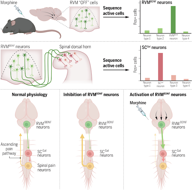
We used several recent technological advances, including the dual activity–dependent approaches ArcTRAP and CANE, single-nucleus RNA sequencing, computational analyses, monosynaptic tracing, and behavioral assays of neuronal activity. We found that the RVM consists of multiple molecular types of γ-aminobutyric acid–mediated (GABAergic) neurons as well as a few types of glutamatergic and serotonergic neurons. Among these, morphine activated a select set of neurons, which together formed a “morphine ensemble.” Synthetic activation of the genetically captured morphine ensemble produced mechanical pain relief, mimicking the effects of morphine, and its inactivation completely abolished the effects of morphine on pain.
Among the neurons in the ensemble, glutamatergic neurons projecting to the spinal cord called RVMBDNF neurons (BDNF, brain-derived neurotrophic factor) were essential. Within the spinal cord, RVMBDNF neurons were connected to GABAergic inhibitory neurons expressing the neuropeptide galanin (SCGal neurons). Inhibition of SCGal neurons completely prevented pain inhibition after administration of morphine or synthetic activation of the RVMBDNF neurons. Notably, the neurotrophic factor BDNF produced within the RVMBDNF neurons was necessary for morphine to have any effect on the sensitivity to painful stimuli. Conversely, increasing BDNF expression in the RVMBDNF neurons markedly potentiated morphine’s effects, with efficacy at doses where morphine alone was insufficient.
CONCLUSION
We discovered that neural activity alone in the RVM induces the key features of morphine-induced mechanical pain relief, and when this activity is inhibited, morphine has little effect. Pain relief is mediated by glutamatergic RVMBDNF neurons projecting to inhibitory SCGal neurons, which attenuate incoming pain signaling in the spinal cord on the way to the brain. Within this circuit, BDNF is an essential component modulating neurotransmission. Finding the molecular identity of neurons that regulate morphine-induced mechanical antinociception advances the search for alternative therapeutic strategies to provide pain relief across various pain conditions."Opioid circuit opens path to pain relief (no public access) "Manipulation of neural circuits targeted by morphine enables pain relief without opioids"
Morphine-responsive neurons that regulate mechanical antinociception (no public access)
Identification of neurons that regulate morphine antinociception.
No comments:
Post a Comment