Showing posts with label organ on a chip. Show all posts

Sunday, January 04, 2026

First breathing lung-on-chip developed using genetically identical cells

Amazing stuff!

"Researchers at the Francis Crick Institute and AlveoliX have developed the first human lung-on-chip model using stem cells taken from only one person. These chips simulate breathing motions and lung disease in an individual, holding promise for testing treatments for infections like tuberculosis (TB) and delivering personalized medicine. ...

Until now, these lung-on-chip devices have been made of a mixture of patient-derived and commercially available cells, meaning they can't fully replicate the lung function or disease progression of a single individual.

In the study, the team ... developed a new lung-on-chip model that contains only genetically identical cells derived from stem cells from a single donor. ...

To further simulate the human lung, AlveoliX has designed specialized machines to impose rhythmic three-dimensional stretching forces on the recreated air sac barrier, mimicking the motion of breathing. This stimulates the formation of microvilli, a key feature of alveolar epithelial cells, to increase surface area for lung functions ..."

"... Tuberculosis (TB) is a very slow-progressing disease in humans, taking months for a person to develop symptoms after being infected with the bacteria Mycobacterium tuberculosis. ... this highlights an increasing need to understand what’s happening in the early stages, when TB bacteria first encounter the body’s defences in the air sacs in the lungs. ..."

From the abstract (emphasis added):
"Immunocompetent and experimentally accessible alveolar systems to study human respiratory diseases are lacking.
Here, we developed a single-donor human induced pluripotent stem cell-derived lung-on-chip (iLoC) containing type II and I alveolar epithelial cells, vascular endothelial cells, and macrophages in a microfluidic device that mimic lung three-dimensional mechanical stretching and air-liquid interface.
Imaging and single-cell RNA sequencing analysis revealed that the iLoC recapitulated cellular profiles present in the human distal lung.
Infection of the iLoC with the human pathogen Mycobacterium tuberculosis (Mtb) showed that both macrophages and epithelial cells were infected but not permissive to bacterial replication. Stochastically, large macrophage clusters containing necrotic macrophages supporting Mtb replication were observed. A genetically engineered autophagy-deficient iLoC revealed that after Mtb infection, macrophage necrosis was higher upon ATG14 deficiency without bacterial replication. Together, we report an autologous, genetically tractable human alveolar model to study lung diseases and therapies."

First breathing 'lung-on-chip' developed using genetically identical cells

Built to breathe: mini ‘lungs’ recreate individual response to infection (original news release) "Lung-on-chip device exposes earliest stages of tuberculosis infection, and opens doors to investigate diversity in disease progression and personalised treatment. "


Image of the lung-on-chip, with cell nuclei in blue, macrophage in magenta, endothelial cells in yellow and tight junctions between epithelial and endothelial cells in white.


Image of a lung-on-chip infected by Mycobacterium tuberculosis, with cell nuclei in blue, dead macrophage in magenta, tight junctions between epithelial and endothelial cells in yellow and TB bacteria in white.


Fig. 1. iPSC-derived iAT2 and iAT1s are differentiated in a lung-on-chip microfluidic device.


Fig. 5. The iLoC mimic early stages of infection with Mtb.


Friday, September 26, 2025

Key driver of a very deadly form of pancreatic cancer spread identified

Good news! Cancer is history (soon)!

"A Cornell-led study has revealed how a deadly form of pancreatic cancer enters the bloodstream, solving a long-standing mystery of how the disease spreads and identifying a promising target for therapy.

Pancreatic ductal adenocarcinoma is among the most lethal cancers, with fewer than 10% of patients surviving five years after diagnosis. ...

New research ... reveals that a biological receptor called ALK7 is responsible, by activating two interconnected pathways that work in tandem. One makes cancer cells more mobile through a process called epithelial-mesenchymal transition, and the other produces enzymes that physically break down the blood vessel walls. ...

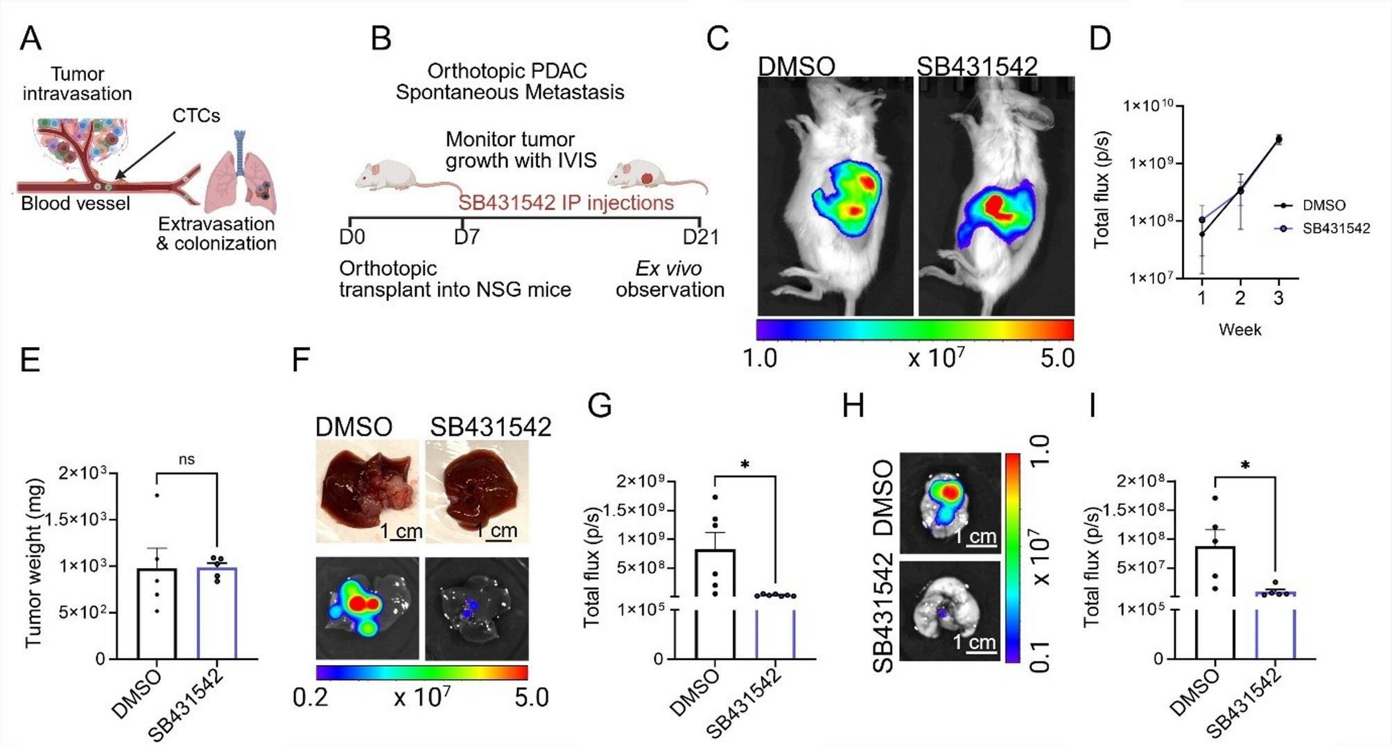
The research helps resolve conflicting findings about ALK7, which some studies had linked to blocking cancer spread while others had tied to driving it. Using mouse models of pancreatic cancer and advanced organ-on-chip systems that mimic human blood vessels, the researchers showed that blocking ALK7 significantly slowed metastasis.

The organ-on-chip system, developed in Lee’s lab, simulates the tumor microenvironment and is superior to animal models for studying different stages of the cancer. Using it, the researchers studied whether ALK7 drives the initial invasion of blood vessels or the later stage, when circulating tumor cells exit the bloodstream to form new tumors in organs such as the lungs or liver. What they found was that cancer cells couldn’t enter blood vessels when ALK7 was inhibited. But when they mimicked a later stage of cancer by placing the cells inside the vessels, they spread quickly, indicating that the timing for treatment is crucial. ..."

From the abstract:
"Breaching the vascular barrier is a critical step in pancreatic ductal adenocarcinoma (PDAC) metastasis, yet the mechanisms enabling this process remain incompletely understood.
Transforming growth factor beta (TGFβ) receptors have been extensively studied in many cancer types. 
However, activin receptor-like kinase 7 (ALK7), one of the TGFβ receptors, is under-investigated, and its roles in PDAC metastasis have been unclear.
This study identifies two distinct but interconnected ALK7-driven non-canonical pathways that promote PDAC dissemination.
The ALK7–β-catenin–EMT axis enhances intrinsic tumor cell motility, driving epithelial-mesenchymal transition (EMT).
In parallel, the ALK7–β-catenin–MMP axis facilitates metastatic invasion by upregulating MMP production, leading to ECM degradation and invadosome formation, which promote vascular barrier breakdown and intravasation.
An orthotopic PDAC metastasis model reveals that both pharmacological and genetic ALK7 inhibition suppresses metastasis.
3D microfluidic vessel-on-chip platforms further demonstrate that ALK7 inhibition preserves basement membrane (BM) integrity, limiting intravasation. While MMP inhibition effectively blocks BM breakdown and intravasation, extravasation remains unaffected, highlighting distinct molecular requirements for different metastatic stages.
These findings establish ALK7 as a dual-function pro-metastatic regulator that orchestrates both tumor cell plasticity and ECM remodeling, positioning ALK7 inhibition as a promising strategy to target early metastatic dissemination in PDAC."

Key driver of pancreatic cancer spread identified | Cornell Chronicle


Fig. 1 Pharmacological ALK7 blockade inhibits spontaneous PDAC metastasis in an orthotopic PDAC model in vivo. 


Friday, September 19, 2025

Wyss Institute received funding to develop first Human Organ Chip model for heavy menstrual bleeding

Good news! It is actually surprising and shocking to learn that: (1) so many women (about 1 in 3 of reproductive age) suffer from heavy menstrual bleeding and (2) that at the end of the first quarter of the 21st century it appears so little can be done to treat it.

Feminists would argue the situation is largely due to the fact that medicine and medical research was dominated by men for centuries and that women bear the heavy menstrual bleeding as a personal, intimate matter.

"The Wyss will use its pioneering Organ Chip technology to create the first human model of heavy menstrual bleeding (HMB), a condition that affects one in three women of reproductive age yet remains underdiagnosed, undertreated, and poorly understood.

The new Organ Chip model will allow researchers to uncover the causal factors and early biomarkers of HMB progression and identify potential non-hormonal therapeutics by combining multi-omics analysis with artificial intelligence (AI)-driven computational approaches. This breakthrough aims to shorten the time women wait for effective treatment from an average of five years to just five months. ...

HMB is more common than asthma or diabetes, yet it has been largely ignored by healthcare systems worldwide. Its consequences are profound. As outlined on Wellcome Leap’s website:

  • Every minute in the U.S., a woman requires a blood transfusion due to menstruation.
  • Up to 50% of reproductive-aged women globally (950 million people) are iron deficient, with chronic HMB expected to be a major contributor.
  • Women with HMB miss an average of 3.6 working weeks annually, costing the U.S. economy more than $94 billion each year.
  • Mental health impacts are significant, with women suffering from HMB experiencing rates of anxiety and depression three times higher than the general female population.
..."

Wyss Institute receives Wellcome Leap funding to develop first Human Organ Chip model for heavy menstrual bleeding "Breakthrough aims to shorten the time women wait for effective treatment from an average of five years to just five months"

Sunday, June 09, 2024

Researchers develop the first-ever complete central nervous system on a chip

Amazing stuff! When will we have a brain on a chip?

"...morphogens, molecules that are manufactured at specific times and places within the embryo and dispersed to dictate the location and shape of our organs. The varying concentrations of morphogens serve as a map that guides the stem cells to their destination and destiny.

Morphogen concentration maps are key to all technologies aimed at making organoids, the laboratory-manufactured miniature versions of living organs that in the past decade have taken over the world of developmental biology. But until now, most researchers have been producing organoids using uniform concentrations of morphogens in petri dishes, which limited them to growing small sections of an organ in each dish, rather than generating a single, miniature version of the complete organ. Now, however, researchers ... have created a miniature version of the entire embryonic central nervous system, from the brain to the bottom of the spinal cord, using a microfluidic chip that mimics the dispersal of the morphogens during embryonic development. ...
The researchers note that their chip does not emulate the earliest stages of the central nervous system’s development. ..."

From the abstract:
"The human nervous system is a highly complex but organized organ. The foundation of its complexity and organization is laid down during regional patterning of the neural tube, the embryonic precursor to the human nervous system. Historically, studies of neural tube patterning have relied on animal models to uncover underlying principles. Recently, models of neurodevelopment based on human pluripotent stem cells, including neural organoids and bioengineered neural tube development models have emerged. However, such models fail to recapitulate neural patterning along both rostral–caudal and dorsal–ventral axes in a three-dimensional tubular geometry, a hallmark of neural tube development. Here we report a human pluripotent stem cell-based, microfluidic neural tube-like structure, the development of which recapitulates several crucial aspects of neural patterning in brain and spinal cord regions and along rostral–caudal and dorsal–ventral axes. This structure was utilized for studying neuronal lineage development, which revealed pre-patterning of axial identities of neural crest progenitors and functional roles of neuromesodermal progenitors and the caudal gene CDX2 in spinal cord and trunk neural crest development. We further developed dorsal–ventral patterned microfluidic forebrain-like structures with spatially segregated dorsal and ventral regions and layered apicobasal cellular organizations that mimic development of the human forebrain pallium and subpallium, respectively. Together, these microfluidics-based neurodevelopment models provide three-dimensional lumenal tissue architectures with in vivo-like spatiotemporal cell differentiation and organization, which will facilitate the study of human neurodevelopment and disease."

From Tip to Tail - Life Sciences | Weizmann Wonder Wander - News, Features and Discoveries Researchers from the University of Michigan and the Weizmann Institute develop the first-ever complete central nervous system on a chip; it faithfully emulates that of a human embryo, from the forebrain to the bottom of the spinal cord


Researchers used color-coded proteins to reveal the identity of the cells in the organoids they produced. In four organoids that simulate the embryo’s central nervous system, the magenta marks proteins associated with the development of the forebrain and the midbrain, the green, the hindbrain and the red, the center of the spinal cord

Wednesday, June 05, 2024

Organ-on-a-chip blood vessels reveal how snake venom destroys capillaries

Good news! Amazing stuff! 

Besides bloodsuckers, snakes might be another species where humans may want to expedite extinction in nature.

"Snakebites kill more than a hundred thousand people every year and maim countless more. ...
The capillary mimics they developed are the latest in ‘organ-on-a-chip’ technology. In a nutshell, cells are grown in special microfluidic devices to create 3D tubes that behave like small blood vessels. Researchers could then pump venom-laced fluid through these mini-tubes and watch what happens to the cells. And it wasn’t pretty: High doses of venom caused entire chunks of vessel wall to die and wither away. ..."

From the abstract:
"Snakebite envenomation is a major public health issue which causes severe morbidity and mortality, affecting millions of people annually. Of a diverse range of clinical manifestations, local and systemic haemorrhage are of particular relevance, as this may result in ischemia, organ failure and even cardiovascular shock. Thus far, in vitro studies have failed to recapitulate the haemorrhagic effects observed in vivo. Here, we present an organ-on-a-chip approach to investigate the effects of four different snake venoms on a perfused microfluidic blood vessel model. We assess the effect of the venoms of four snake species on epithelial barrier function, cell viability, and contraction/delamination. Our findings reveal two different mechanisms by which the microvasculature is being affected, either by disruption of the endothelial cell membrane or by delamination of the endothelial cell monolayer from its matrix. The use of our blood vessel model may shed light on the key mechanisms by which tissue-damaging venoms exert their effects on the capillary vessels, which could be helpful for the development of effective treatments against snakebites."

ScienceAdvisor


Figure 7 Schematic overview showing the mechanisms by which tissue-damaging toxins exert their effects


Thursday, April 18, 2024

Understanding the 3D Cell Culture Revolution

Recommendable! Seems to be a nice, brief overview article!
Caveat: I only skimmed this article.

Understanding the 3D Cell Culture Revolution | The Scientist Magazine® 3D cell culture techniques closely mimic in vivo conditions, generating more accurate data for disease modeling and drug toxicity testing.

3D Cell Culture Model Types
3D cell culture types include scaffold-free spheroids, scaffold-based organoids, and microfluidic-based organ on a chip devices. These are useful in disease research applications, such as studying the brain tumor microenvironment, modeling inflammatory conditions like chronic obstructive pulmonary disease (COPD), and investigating drug toxicity in vitro.


Tuesday, December 19, 2023

A malaria drug treatment could save babies’ lives regardless whether pregnant mother contracted malaria or not

Good news! What a surprise!

"... To try to reduce the risk of malarial infection, the World Health Organization recommends that pregnant women in low-income countries be treated with a combination of the antimalarial drugs sulfadoxine and pyrimethamine (SP). Curiously, a recent study found that this treatment also seemed to increase the birth weight of treated mothers’ babies, regardless of whether they contracted malaria. ..."

A malaria drug treatment could save babies’ lives Human organ chip research shows that a common antimalarial combination could reverse the negative effects of malnutrition in the female digestive tract that lead to low birth weight infants

Intermittent preventive therapy for malaria during pregnancy using 2 vs 3 or more doses of sulfadoxine-pyrimethamine and risk of low birth weight in Africa: systematic review and meta-analysis (open access)

The Intestine Chip was created by taking healthy intestinal cells donated by female patients and culturing them inside a human Organ Chip device. This process allowed the team to create the first in vitro model of the adult female intestine and replicate the hallmarks of malnutrition to find treatments. 


Monday, November 21, 2022

Placenta-on-a-chip simulates nutrient transport between mother and foetus

When will we have a human on a chip? 😊

"Researchers ... have developed a “placenta-on-a-chip” that closely mimics the molecular exchange of nutrients between mother and foetus during pregnancy. ...  created the device using a pair of microfluidic channels, separated by an intricate network of hydrated fibres cultured on each side with different placental cells. The setup enabled the team to recreate disruptions to nutrient exchange caused by placental malaria, and could be a key step towards developing a treatment for the disease. ..."

From the abstract:
"... In this work, a placenta-on-a-chip model is developed to mimic the nutrient exchange between the fetus and mother under the influence of PM [placenta malaria]. In this model, trophoblasts cells (facing infected or uninfected blood simulating maternal blood and termed “trophoblast side”) and human umbilical vein endothelial cells (facing uninfected blood simulating fetal blood and termed “endothelial” side) are cultured on the opposite sides of an extracellular matrix gel in a compartmental microfluidic system, forming a physiological barrier between the co-flow tubular structure to mimic a simplified maternal–fetal interface in placental villi. The influences of infected erythrocytes (IEs) sequestration through cytoadhesion to chondroitin sulfate A (CSA) expressed on the surface of trophoblast cells, a critical feature of PM, on glucose transfer efficiency across the placental barrier was studied. ... The results demonstrated that CSA-binding IEs added resistance to the simulated placental barrier for glucose perfusion and decreased the glucose transfer across this barrier. The results of this study can be used for better understanding of PM pathology and development of models useful in studying potential treatment of PM."

Placenta-on-a-chip simulates nutrient transport between mother and foetus – Physics World

Sunday, July 31, 2022

Organ-on-a-chip system replicates rare neuromuscular disorders, finds new treatment without animal testing

Good news! This could be a breakthrough for organ on a chip systems to develop new drugs!

"Microphysiological systems, also known as organ-on-a-chip systems, are able to replicate key aspects of the human body and its function. These devices can be used with various living human cells to accelerate drug development, disease modeling, and personalize medicine — and the field is growing fast.
Now, researchers in the United States have used an advanced microphysiological system that mimics the pathology of chronic inflammatory demyelinating polyneuropathy and multifocal motor neuropathy, two very rare but very devastating neuromuscular diseases that cannot be replicated in animal models like rodents or primates. ...
There are over 7,000 rare diseases with no effective treatments, and only about 400 are being actively researched due to a variety of reasons, including the lack of animal models. Moreover, about 9 in 10 drug candidates that show promising results in tests on rodents eventually fail in clinical trials on humans. ..."

From the abstract:
"Chronic autoimmune demyelinating neuropathies are a group of rare neuromuscular disorders with complex, poorly characterized etiology. Here the authors describe a phenotypic, human-on-a-chip (HoaC) electrical conduction model of two rare autoimmune demyelinating neuropathies, chronic inflammatory demyelinating polyneuropathy (CIDP) and multifocal motor neuropathy (MMN), and explore the efficacy of TNT005, a monoclonal antibody inhibitor of the classical complement pathway. Patient sera is shown to contain anti-GM1 IgM and IgG antibodies capable of binding to human primary Schwann cells and induced pluripotent stem cell-derived motoneurons (MNs). Patient autoantibody binding is sufficient to activate the classical complement pathway, resulting in detection of C3b and C5b-9. A HoaC model, using a microelectrode array with directed axonal outgrowth over the electrodes treated with patient sera, exhibits reductions in MN action potential frequency and conduction velocity. TNT005 rescued the serum-induced complement deposition and functional deficits while treatment with an isotype control antibody has no rescue effect. These data indicate that complement activation by CIDP and MMN patient serum is sufficient to mimic neurophysiological features of each disease and that complement inhibition with TNT005 is sufficient to rescue these pathological effects and provide efficacy data included in an investigational new drug application, demonstrating the model's translational potential."

Human-on-a-chip system replicates rare neuromuscular disorders, finds new treatment without animal testing The FDA approved the first clinical trial using efficacy data collected from a microphysiological system. Here's why that's a pretty big deal.


Sunday, April 10, 2022

Fighting viruses is as easy as breathing on a human lung chip

Amazing stuff! What is all the RAGE about? Hint: Receptor for advanced glycation end products. 😄
Or how to "Creating a flu-on-a-chip".

"The average person will take more than 600 million breaths over the course of their life. ... Now, new research ... has revealed that this constant pattern of stretching and relaxing [of the lungs] does even more – it generates immune responses against invading viruses. ...
Using a Human Lung Chip that replicates the structures and functions of the lung air sac, or “alveolus,” the research team discovered that applying mechanical forces that mimic breathing motions suppresses influenza virus replication by activating protective innate immune responses. They also identified several drugs that reduced the production of inflammatory cytokines in infected Alveolus Chips, which could be useful in treating excessive inflammation in the lung. ...
Human Organ Chips were developed to address this problem, and have been shown to faithfully replicate the functions of many different human organs in the lab, including the lung. ... since 2017, ... researchers have been working on replicating various diseases in Lung Airway and Alveolus Chips to study how lung tissues react to respiratory viruses that have pandemic potential and test potential treatments. ..."

From the abstract:
"Mechanical breathing motions have a fundamental function in lung development and disease, but little is known about how they contribute to host innate immunity. Here we use a human lung alveolus chip that experiences cyclic breathing-like deformations to investigate whether physical forces influence innate immune responses to viral infection. Influenza H3N2 infection of mechanically active chips induces a cascade of host responses including increased lung permeability, apoptosis, cell regeneration, cytokines production, and recruitment of circulating immune cells. Comparison with static chips reveals that breathing motions suppress viral replication by activating protective innate immune responses in epithelial and endothelial cells, which are mediated in part through activation of the mechanosensitive ion channel TRPV4 and signaling via receptor for advanced glycation end products (RAGE). RAGE inhibitors suppress cytokines induction, while TRPV4 inhibition attenuates both inflammation and viral burden, in infected chips with breathing motions. Therefore, TRPV4 and RAGE may serve as new targets for therapeutic intervention in patients infected with influenza and other potential pandemic viruses that cause life-threatening lung inflammation."

Fighting viruses is as easy as breathing Human Lung Chip reveals the effects of breathing motions on lung immune responses and leads to repurposing of potential therapeutics for respiratory diseases, including COVID-19

Thursday, May 06, 2021

Human organ chips enable rapid drug repurposing for COVID-19

Recommendable! Amazing stuff! Microfluidics in action!

"A Wyss Institute-led collaboration spanning four research labs and hundreds of miles has used the institute’s organ-on-a-chip (Organ Chip) technology to identify the antimalarial drug amodiaquine as a potent inhibitor of infection with SARS-CoV-2, the virus that causes COVID-19.  ...
When tested in their more sophisticated microfluidic Lung Airway Chip, which had been infected with a pseudotyped SARS-CoV-2 virus, they found that most of these drugs, including hydroxychloroquine and chloroquine, were not effective. However, another antimalarial drug, amodiaquine, was highly effective at preventing viral entry. ...
The human Airway Chip that the Wyss team developed for these studies is a microfluidic device about the size of a USB memory stick that contains two parallel channels separated by a porous membrane. Human lung airway cells are grown in one channel that is perfused with air, while human blood vessel cells are grown in the other channel, which is perfused with liquid culture medium to mimic blood flow. Cells grown in this device naturally differentiate into multiple airway-specific cell types in proportions that are similar to those in the human airway, and develop traits observed in living lungs such as cilia and the ability to produce and move mucus. Airway Chip cells also have higher levels of angiotensin-converting enzyme-2 (ACE2) receptor protein, which plays a central role in lung physiology and is used by SARS-CoV-2 to infect cells."

Human organ chips enable rapid drug repurposing for COVID-19 – Harvard Gazette Emulating human lung airway in vitro identified helpful effects of antimalarial drug amodiaquine, now in clinical trials

Here is the underlying research article (dated 4/15/2020):

Sunday, February 16, 2020

Bone marrow-on-a-chip models damage and disease

Recently, I blogged here about body-on-a-chip!  Now, a new organ-on-a chip has been reported! Amazing stuff!

"Until David Chou, Viktoras Frismantas and their colleagues developed the bone marrow chip, the only way to study living marrow tissue effectively was to take painful biopsies from the bones. ... we are in a position to assist in the design of human clinical trials for rare genetic disorders and advance personalized medicine in ways not possible before"

Bone marrow-on-a-chip models damage and disease – Physics World: Human bone marrow chip provides a reliable model to study bone marrow damage by radiation, drugs and disease

Thursday, February 06, 2020

TAU, Harvard Researchers Build Human 'Body-On-Chips' To Better Predict Drug Response

Amazing stuff! Up to 10 organ-on-chip were fluidically linked together to form a a functional human Body-on-Chips platform! 

"The interrogator [body-on-chips platform] enabled the team to culture, perfuse, and link the living human cultured tissues in a multi-Organ Chip system, as well as add and sample the medium in a fully programmable way using the device’s robotic liquid transfer capabilities, while continuously monitoring tissue integrity ..."

This should promote and boost diagnostics, drug testing and so much more ...

TAU, Harvard Researchers Build Human 'Body-On-Chips' To Better Predict Drug Response | News Briefs: A team of over 50 scientists created and linked up eight organs-on-chips, including kidney, liver, brain, and heart, to pave the way for improved clinical testing. | NoCamels

Sunday, July 21, 2019

Human Gut Microbiome On A Chip

Posted: 7/21/2019

The Wyss Institute at Harvard University may have delivered a momentous breakthrough by developing a very realistic, viable human gut microbiome on a chip. If confirmed, this has great potential for further and rapid medical progress!

“A research team ... has developed a ... ‘organ-on-a-chip’ (Organ Chip) microfluidic culture technology. ... now able to culture a stable complex human microbiome in direct contact with a vascularized human intestinal epithelium for at least five days in a human Intestine Chip in which an oxygen gradient is established that provides high levels to the endothelium and epithelium while maintaining hypoxic conditions in the intestinal lumen inhabited by the commensal bacteria. Their “anaerobic Intestine Chip” stably maintained a microbial diversity similar to that in human feces over days and a protective physiological barrier that was formed by human intestinal tissue. The study is published in Nature Biomedical Engineering.” (S1; emphasis added)

Sources (S):