Showing posts with label morphogenesis. Show all posts

Friday, September 04, 2026

Wrinkled or smooth? Cell growth mechanics determine shape of plant organs

Amazing stuff!

"When inner and outer cell layers of sepals – the leaf-like plant organs that cover and protect the flower bud before it opens – all grow upward, the organs stay smooth and uniformly stiff, which is optimal for the plant, according to a new study. 

The study ... compared a wild type Arabidopsis plant with a mutant one that grew with wrinkled sepals. For the first time, the researchers were able to image both the inner and outer layers of the sepal to understand factors that controlled smoothness and wrinkles. ..."

From the highlights and abstract:
"Highlights
• Smooth sepal formation requires aligned growth directions and comparable stiffness
• Ectopic expression of AS2 causes buckling of the outer epidermis
• Buckles form due to misaligned growth directions and unequal epidermal stiffness
• Buckling promotes the PIN1 convergence to initiate outgrowths

Summary
Nature exhibits organs of various shapes and forms, ranging from smooth to undulated morphologies.
How cells coordinate their growth to produce smoothly shaped leaves and leaf-like organs such as sepals remains unclear.
We identified a mutant, as2-7D, that exhibits ectopic expression of ASYMMETRIC LEAVES 2 (AS2) on the outer epidermis.
Our analysis reveals that ectopic AS2 expression causes the outer epidermis of as2-7D sepals to buckle during early stages of sepal development.
Buckling is caused by conflicting cell growth directions and unequal tissue stiffness across the epidermal layers.
Overexpression of cyclin-dependent kinase (CDK) inhibitor Kip-related protein 1 (KRP1) in as2-7D aligns the growth directions of the outer epidermal cells along the longitudinal axis, increases the overall stiffness of the outer epidermis, and restores sepal smoothness.
Further experiments suggest that buckling promotes the convergence of auxin efflux transporter protein PIN-FORMED 1 (PIN1) to initiate pointed outgrowths. Thus, we show that growth along the longitudinal axis during early developmental stages and comparable stiffness across both epidermal layers of Arabidopsis thaliana sepals are essential for smoothness, as seen in the wild type.
Our findings suggest that in addition to molecular cues influencing tissue mechanics, tissue mechanics can also modulate molecular signals, giving rise to well-defined shapes."

Wrinkled or smooth? Cell growth mechanics determine shape of plant organs | Cornell Chronicle


Graphical abstract


Figure 1. The ectopic expression of AS2 on the outer epidermal layer disrupts sepal smoothness in as2-7D mutant


Monday, January 12, 2026

Broken bone: The same nerves that transmit the feeling of pain later enter a pro-regenerative state to heal the broken bone

Amazing stuff!

"... Breaking a bone hurts because sensory neurons are triggered by the trauma, and they signal pain to the brain. But that’s not these neurons’ only job, researchers discovered: They also step in to lead bone repair.

To discover this, researchers zoomed in on the nerves that penetrate into bone in mice, tracing them all the way back to the central nervous system, as well as sequencing individual neurons before and after fractures. “We created the first comprehensive single-cell atlas of bone-innervating sensory neurons,”  ... This revealed that the same nerves that transmit the feeling of pain later enter what the team calls a “pro-regenerative state … they produce and release proteins that promote the generation of new neurons, blood vessels, and of course, bone and cartilage.” Of particular importance is a protein called fibroblast growth factor 9 (FGF9), which is a key coordinator of bone repair, guiding stem cells to transform into bone-building cells. When the researchers removed the nerves that produce FGF9 from mice, their bones struggled to heal. ..."

From the abstract of the Perspective:
"The skeleton has a remarkable ability to grow: It greatly expands in size from birth to adulthood and continues to remodel throughout adult life, completely replacing itself every 10 years. 
But it is also able to regenerate after injury, efficiently regrowing lost bone. Bone injuries are common, with an estimated 6 million to 10 million fractures occurring each year in the United States.
A lack of efficient healing can result in long-term disability, and in the geriatric population, complications from fractures are associated with increased morbidity and mortality.
Because bone repair reflects the successful integration of multiple cellular and molecular processes, it is challenging to precisely define the signaling hierarchy that produces optimal healing. ... report an unexpected role for sensory nerves in bone healing, providing insights into communication between the nervous system and the cells responsible for bone repair."

From the editor's summary and abstract:
"Editor’s summary
Sensory neurons innervating bones can modulate the healing process (bone regeneration) after a fracture. Xu et al. used single-cell transcriptomics to characterize the bone-innervating dorsal root ganglia (DRG) neurons before and after an ulnar stress fracture in mice ... The experiments revealed temporally dynamic responses of the DRG neurons after the fracture. Moreover, the authors identified fibroblast growth factor 9, released by sensory neurons, as a major neural regulator in bone regeneration after injury. These findings show that pathways activated by bone-innervating neurons after an injury might be exploited for accelerating bone fracture repair. ...

Structured Abstract
INTRODUCTION
The profound pain accompanying bone fracture is mediated by somatosensory neurons, which also appear to be required to initiate bone regeneration. Primary somatosensory neurons comprise a diverse subset of neurons, which communicate information about the external environment and internal state to the central nervous system, enabling perception and reaction to a wide range of stimuli including pain.

RATIONALE
Most work in skeletal neurobiology has focused on understanding bone nociceptive pathways, but recent studies provide evidence that sensory nerves also function to initiate bone formation during skeletal morphogenesis. To what extent such bone morphogenic and nociceptive actions are mediated by distinct neuronal pathways has been difficult to study in part due to lack of tractable model systems for studying nerve-bone interactions and the extensive heterogeneity of peripheral sensory neurons.

RESULTS
To characterize neuroanatomical circuitry mediating skeletal nociception and regeneration, dorsal root ganglia (DRG) neurons innervating murine long bones were profiled by retrograde nerve labeling and single-cell transcriptomics before and after experimental fracture.
Highest labeling across CGRP+ and Aβ-Field LTMR neurons was identified, which have nociceptive or mechanoreceptive functions.
Dynamic changes associated with sensory neuron response to injury reflected the phasic nature of bone repair.
At early time points, DRG neurons showed signatures of pain perception and inflammatory responses.
At later time points, DRG neurons demonstrated transcriptomic changes more characteristic of a regenerative response, including mitogenic, angiogenic and osteogenic signals.
This includes expression of morphogens in the fracture reparative phase such as Tgfb1, Fgf9, and Shh.
Two methods to surgically or genetically denervate fractured bones were used to implicate defective mesenchymal cell proliferation and osteo differentiation as underlying poor bone repair with loss of innervation.
Finally, multitissue single-cell RNA-sequencing and interactome analyses implicated neuron-derived fibroblast growth factor 9 (FGF9) as a potent regulator of fracture repair, confirmed by in vivo sensory nerve–specific ablation studies.

CONCLUSION
In sum, somatosensory neurons innervating bone are a group of neurons with nociceptive and mechanoreceptive functions, which are transcriptionally responsive to bone injury in a temporal dynamic fashion and positively regulate fracture healing through FGF9-FGFR signaling."

ScienceAdviser

Not just a pain in the bone (Perspective, no public access) "Growth factors secreted by sensory nerves promote fracture healing"


Fig. 2 Temporal responses of sensory neurons to fracture injury


When a bone is broken, sensory nerves alert the brain about the painful trauma. But after they’ve reported the break, they go into repair mode, producing FGF9 (red dots)—which, among other important messages, tells special stem cells to start the process of becoming bone cells (osteoblasts).


Tuesday, December 30, 2025

Detailed cell map unlocks secrets of how reproductive organs form during prenatal development

Good news! Amazing stuff!

"New research has mapped the cell types that specialise to form reproductive organs in both sexes, identifying key genes and signals that drive this process. The findings offer important insights into conditions affecting the reproductive organs, and how environmental chemicals may affect reproductive health.

Researchers ... used a combination of single-cell and spatial genomics technologies to analyse over half a million individual human cells from the developing reproductive system.  ..."

From the abstract:
"The human reproductive tract is essential for species perpetuation and overall health. Its development involves complex processes of sex specification, tissue patterning and morphogenesis, the disruption of which can cause lifelong issues, including infertility.
Here we present an extensive single-cell and spatial multi-omic atlas of the human reproductive tract during prenatal development to provide insights beyond those that are possible with smaller-scale, organ-focused studies.
We describe potential regulators of sexual dimorphism in reproductive organs and pinpoint previously unknown genes involved in Müllerian duct emergence and regression and urethral canalization of the penis.
By combining histological features with gene expression and chromatin accessibility data, we define transcription factors and signalling events potentially involved in the regionalization of the Müllerian and Wolffian ducts. 
We also refine how the HOX code is established in distinct reproductive organs and reveal that the expression of thoracic HOX genes is increased in the rostral mesenchyme of the fallopian tube and epididymis.
Our findings further indicate that epithelial regionalization of the fallopian tube and epididymis, which probably contribute to sperm maturation and capacitation, is established during development.
By contrast, later events are necessary for regionalization of the uterocervical canal epithelium.
Finally, on the basis of single-cell data and fetal-derived organoids, we show that the fetal uterine epithelium is vulnerable to oestrogen-mimicking endocrine disruptors.
By mapping sex-specific reproductive tract regionalization and differentiation at the cellular level, our study provides valuable insights into causes and potential treatments of developmental reproductive disorders."

Detailed cell map unlocks secrets of how reproductive organs form "Scientists chart a comprehensive cell atlas of the developing human reproductive system, offering new clues into congenital disorders."


Fig. 1: Single-cell resolved spatiotemporal atlas.


Saturday, August 30, 2025

Scientists debut a new foundational atlas of the plant life cycle

Good news!

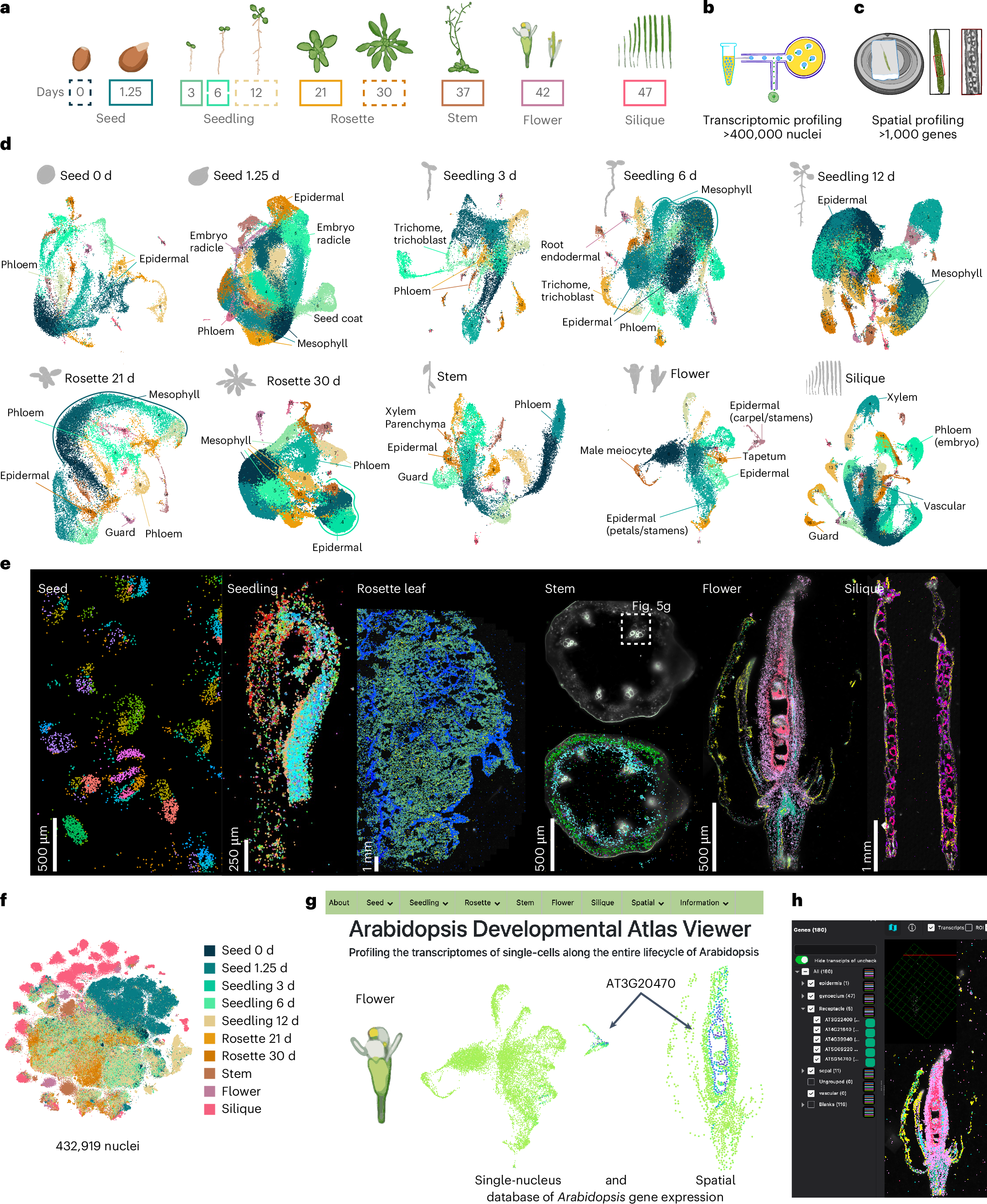
"... researchers have now established the first genetic atlas to span the entire Arabidopsis life cycle. The new atlas—created using detailed single-cell and spatial transcriptomics—captures the gene expression patterns of 400,000 cells within multiple developmental stages as Arabidopsis grows from a single seed to a mature plant. The publicly available resource will be hugely informative to future studies of different plant cell types and developmental stages, and how they respond to stress and environmental stimuli. ..."

From the abstract:
"Arabidopsis has been pivotal in uncovering fundamental principles of plant biology, yet a comprehensive, high-resolution understanding of its cellular identities throughout the entire life cycle remains incomplete.
Here we present a single-nucleus and spatial transcriptomic atlas spanning ten developmental stages, encompassing over 400,000 nuclei from all organ systems and tissues—from seeds to developing siliques. Leveraging paired single-nucleus and spatial transcriptomic datasets, we annotate 75% of identified cell clusters, revealing striking molecular diversity in cell types and states across development.
Our integrated approach identified conserved transcriptional signatures among recurrent cell types, organ-specific heterogeneity and previously uncharacterized cell-type-specific markers validated spatially.
Moreover, we uncover dynamic transcriptional programs governing secondary metabolite production and differential growth patterns, exemplified by detailed spatial profiling of the compact yet complex apical hook structure; this profiling revealed transient cellular states linked to developmental progression and hormonal regulation, highlighting the hidden complexity underlying plant morphogenesis.
Functional validation of genes uniquely expressed within specific cell contexts confirmed their essential developmental roles, underscoring the predictive power of our atlas.
Collectively, this comprehensive resource provides an invaluable foundation for exploring cellular differentiation, environmental responses and genetic perturbations at high resolution, advancing our understanding of plant biology."

Scientists debut a new foundational atlas of the plant life cycle - Salk Institute for Biological Studies "Salk Institute researchers map every cell type and developmental state across the entire life cycle of model plant Arabidopsis"


Fig. 1: A spatially resolved transcriptional atlas of the Arabidopsis life cycle.


Tuesday, March 18, 2025

Tunneling nanotube–like structures regulate distant cellular interactions during heart formation

Amazing stuff!

From the perspective abstract:
"The heart is the first organ to become functional during embryonic development in vertebrates. The process begins with the formation of the heart tube, which consists of an inner endothelial lining (endocardium) and a layer of muscle cells (myocardium), separated by a thick extracellular matrix called the cardiac jelly. Subsequent development of the heart ventricles involves the formation of trabeculae—muscular ridges lined by the endocardium. How signals pass between the endocardium and myocardium across the cardiac jelly to ensure normal development is not fully understood. ...
report that, in mice, the myocardium and endocardium of the early heart tube communicate directly through tiny membrane-enclosed projections called tunneling nanotube–like structures (TNTLs). Loss of TNTLs disrupts trabecular formation and thus ventricular development and embryonic viability."

From the editor's summary and abstract:
"Editor’s summary
During cardiac development, heart muscle cells (the myocardium) and inner lining cells (the endocardium) are separated by a space filled with cardiac jelly. ... discovered that despite this physical separation, the two cell types communicate with each other through tiny structures called tunneling nanotube-like microstructures (TNTLs) ... TNTLs extend across the cardiac jelly, allowing direct cell-cell contact, signal transduction, and selective protein transfer. Disrupting TNTLs leads to abnormal heart wall formation, highlighting the importance of TNTLs in cardiac development. ...

Structured Abstract
INTRODUCTION
Heart development is a highly orchestrated process dependent on dynamic interactions between the myocardium and the endocardium. The two layers are separated by a noncellular matrix called cardiac jelly and communicate through signaling pathways involving membrane-bound receptors and ligands. However, the mechanisms enabling such signaling interaction over physical distances remain poorly understood.
In this work, we characterized tunneling nanotube–like structures (TNTLs), which we found physically connecting cardiomyocytes (CMs) in the myocardium to endocardial cells (ECs) in the endocardium. These structures likely help to facilitate long-distance intercellular communication essential for heart formation.

RATIONALE
Heart formation relies on precise signaling interactions between the myocardium and endocardium, particularly during trabecular development. Signaling pathways, such as Notch1, Vegf, and Nrg1, have ligands and receptors segregated across these two cardiac layers. The mechanisms enabling these long-distance interactions across the intervening cardiac jelly are unclear.
We hypothesized that TNTL structures exist between the cardiac layers and could mediate intercellular long-distance communication in the developing heart, allowing for the transport of signaling molecules and cytoplasmic proteins between them.

RESULTS
We used genetic labeling, contact-tracing techniques, and advanced imaging to demonstrate the existence of TNTLs in mouse embryonic hearts. These TNTLs extended from CMs to ECs across the cardiac jelly, establishing direct connections that enable signal transduction and cytoplasmic protein transfer.
The TNTLs were identified in the heart through the genetic labeling of cellular protrusions. During mouse development TNTLs were shown to form between CMs and ECs as early as embryonic day 8.0. The filamentous structures inside TNTL were characterized by three-dimensional imaging and the reconstruction of the electron microscopy (EM) and cryo-EM images.
The TNTLs contained actin filaments, and TNTL formation depended on actin polymerization. The presence of actin filaments in TNTLs was confirmed in a transgenic mouse line that could label actin filaments with a fluorescent marker. Inhibiting actin polymerization chemically or by ablating the small guanosine triphosphatase, Cdc42, eliminated TNTLs.
The TNTLs were involved in regulating Notch1 signaling and other signaling pathways. TNTLs were sufficient to activate Notch1 signaling in ECs, with ligands from CMs transported through these microstructures to ECs.
Loss of TNTLs resulted in reduced Notch signaling and other signaling pathways. TNTLs were able to transport signaling molecules, cytoplasmic proteins, and trafficking vesicles, underscoring their role as conduits for intercellular communication.
The TNTLs were essential in cardiac morphogenesis. Disruption of TNTLs in embryonic hearts resulted in impaired ventricular wall morphogenesis, evidenced by loss of trabeculae and defective myocardial growth.

CONCLUSION
In this work, we identified TNTLs as a critical mechanism for long-distance intercellular communication during heart development. These actin-rich structures physically bridge the myocardium and endocardium, allowing for the efficient exchange of signaling molecules necessary for trabecular formation and ventricular wall morphogenesis.
Disruption of TNTLs compromises these interactions, highlighting their essential role in heart patterning. This work provides insights into mechanisms of cellular communication and suggests that TNTL formation might help cells to regulate long-distance cell-cell communication and modulate tissue patterning in mammalian systems. ..."

Tunneling through cardiac jelly (no public access) "Membrane projections from muscle cells enable signaling in the developing mouse heart"

Tunneling nanotube–like structures regulate distant cellular interactions during heart formation | Science (no public access)


Microstructure regulates signaling interaction and cytoplasmic proteins transfer between CMs and ECs.