Showing posts with label hepatology. Show all posts

Saturday, May 09, 2026

Satellite virus spreads through viral Trojan Horses

Amazing stuff!

"Satellite viruses replicate their genomes within host cells but depend on helper viruses for spread.
Deltavirus, or hepatitis D-like virus, is a hepatitis B satellite virus that causes severe viral hepatitis in humans.
Recently, deltaviruses have been found in many animals outside of the liver, suggesting that their diversity and disease potential are underestimated.
The current paradigm for satellite viruses is that they simply “borrow” envelope proteins from related helper viruses.
McKellar et al. used electron and super-resolution microscopy in rhabdovirus, herpesvirus, and arenavirus systems to show that deltavirus ribonucleoproteins can package themselves within a variety of helper virions. This viral Trojan horse mode of transmission could broaden deltavirus host range and explain overlooked infections in humans."

From the highlights and abstract:
"Highlights
• Deltaviruses hitchhike within helper virus virions, using them as viral Trojan Horses
• The Trojan Horse model is mandatory for productive herpesvirus-deltavirus associations
• This mode of propagation favors deltavirus infectivity
• Trojan Horse model warrants screening for extra-hepatic deltavirus infections in humans

Summary
Hepatitis D-like satellite viruses, known as deltaviruses, have been recently discovered in a wide range of animals. These viruses are thought to expropriate glycoproteins from helper viruses to form infectious particles.
Here, we challenge this paradigm and demonstrate that deltaviruses are packaged within helper virus particles, using them as viral Trojan Horses for cell entry. By leveraging orthogonal electron and optical super-resolution microscopy, we visualize deltaviruses enclosed within virions from rhabdo-, herpes-, and arenavirus families.
We show that this conserved hitchhiking mechanism ensures concomitant deltavirus-helper virus spread, thereby promoting the dissemination of deltaviruses, broadening their host range, and expanding their tropism.
Our findings reveal a previously unrecognized mode of viral transmission, providing a framework to investigate overlooked deltavirus infections outside of the human liver."

In Other Journals | Science


Graphical abstract


Figure 1 A subset of VSV virions is morphologically modified after superinfection of deltavirus-replicating cells


Friday, May 08, 2026

A Journey to the Center of the Liver: A first genetic atlas of a healthy human liver at a resolution of 2 microns

Amazing stuff!

"... In a new study ... present the first genetic atlas of a healthy human liver at a resolution of 2 microns. The findings show that the division of labor in the human liver differs from that of other mammals and is more extensive than previously recognized, helping explain why certain regions of the liver are particularly vulnerable to fatty liver disease. ...

the liver has a remarkable capacity for regeneration, healthy individuals can donate a substantial portion of their livers to patients in need. ..."

From the abstract:
"Reconstructing gene expression atlases for human tissues is challenging due to limited access to healthy samples from live individuals.
Neurologically deceased donors often show ischaemic changes, and tissues near diseased regions may have altered gene expression. The liver, with its unique regenerative capacity, allows analysis from live healthy donors.
Here, using spatial transcriptomics (Visium, Visium HD3, multiplexed error-robust fluorescence in situ hybridization (MERFISH)4 and PhenoCycler imaging) and single-nucleus RNA sequencing, we analysed 16 liver samples: 8 from young live healthy donors and 8 from individuals with liver pathology, sampling ‘adjacent normal’ tissue.
Livers from live healthy donors displayed significant gene expression differences compared with the adjacent normal tissues from individuals with liver pathology.
Hepatocytes and non-parenchymal cells exhibited marked zonation along the porto–central axis of the liver lobules, with key functions being pericentrally shifted compared to mice and other mammals.
Our atlas identified dynamic programmes in early steatotic hepatocytes, including a decline in nuclear-encoded mitochondrial proteins and a compensatory increase in mitochondria-encoded transcripts.
This study presents a spatial gene expression reference for the healthy human liver and insights into hepatocyte changes in early steatosis."

A Journey to the Center of the Liver - Life Sciences | Weizmann Wonder Wander - News, Features and Discoveries "A first-of-its-kind genetic atlas reveals how human liver cells divide their labor – and why some regions are especially vulnerable to fatty liver disease"


Fig. 1: A spatial expression atlas of human and mammalian livers.


Fig. 2: Zonation of hepatocyte gene expression.


Friday, April 17, 2026

Growing engineered liver tissue on demand directly in the body

Good news!

"... “We asked if it would be possible to first implant a small-scale liver construct and then drive it to expand in the body following its engraftment. A sufficiently grown, functional ‘satellite liver’ could immediately relieve the metabolic burden in a damaged liver and help bridge the time until a transplant becomes available,”  ..."

From the abstract:
"Despite the promise of engineered tissue implants for the treatment of organ failure, scaling of these constructs to sizes of therapeutic relevance remains a barrier to clinical translation.
Here, we propose a strategy to circumvent this limitation: to instead implant a small-scale construct and then induce it to grow in situ after its engraftment into a host.
Using engineered liver tissue as a proof-of-concept application, we integrated synthetic biology and tissue engineering tools to build liver tissues that can be expanded on-demand after implantation in vivo.
To achieve this goal, we first identified the combination of Yes-associated protein (YAP) and growth factor (GF) signaling as sufficient to drive human hepatocyte proliferation in dense, three-dimensional engineered tissues.
We then engineered control of these signaling axes using synthetic biology tools to drive human liver tissue expansion both in vitro and in vivo.
As such, this work establishes a genetic strategy for generating large organ implants through bioengineered on-demand outgrowth via synthetic biology triggering (BOOST)."

Growing liver tissue on demand directly in the body "New study combines tissue engineering with synthetic biology tools to grow healthy liver tissue inside the body, and lays foundation for “smart” solid organ therapies"

Fig. 1. GF and YAP signaling synergize to induce proliferation of dense cultures of HEPs.


The genetic “BOOST” strategy, explained in his graphic, integrates tissue engineering and synthetic biology tools to enable on-demand liver growth inside the body. By specifically rewiring the gene expression of primary liver hepatocytes and supportive fibroblast cells, a tissue growth program is switched on in a small, engineered liver construct after its implantation into recipients and upon addition of an inducing agent (shown as a pill). As a result, the hepatocytes in the construct start and continue to proliferate until a desired construct size has been reached and the inducing signal is not provided anymore. In mice, BOOST resulted in robust and healthy liver growth.


Saturday, January 24, 2026

New hope for liver cirrhosis treatment

Good news!

"Chronic liver disease gets worse when the liver's immune system stays overactive, resulting in ongoing inflammation and scarring. Signals from the gut make the situation worse by encouraging immune cells to release harmful molecules. Macrophages and monocytes are central players, intensifying injury and shifting the body's defenses toward a more aggressive state. Among them, liver macrophages are essential and are now being explored as therapeutic targets. ..."

From the highlights and abstract:
"Highlights
• PAF/PAF-R signaling pathway is overactivated in patients with cirrhosis.
• PAF/PAF-R blockade ameliorates liver injury during experimental cirrhosis.
• Antagonizing PAF/PAF-R pathway restores homeostatic Treg/Th17 immune balance.
• Promoter DNA methylation controls PAF-R expression in hepatic macrohages.

Abstract
Background and aims
Platelet-activating factor (PAF) phospholipid is mainly produced by macrophages and involved in pro-inflammatory responses. We evaluated the regulation of PAF-R gene expression during experimental cirrhosis and whether antagonizing its ligand PAF improves liver function and inflammation.

Methods
Patients with cirrhosis and CCl4-induced cirrhotic C57Bl/6 mice were included in the study. A subgroup of mice was treated with either PAF antagonist BN-52021 or a DNMT inhibitor, Aza, for two weeks before laparotomies. Sorted hepatic macrophages were subjected to a genome-wide DNA methylation study, and Ptafr expression analysed by Western Blot, qPCR, and immunohistochemistry in liver tissue. Immortalized Kupffer cells (imKCs) were stimulated with PAF and antigenic ligands. Cytokine and chemokine expression were measured. Biochemical and hepatic markers of liver damage were assessed.

Results
Hepatic PAF-R increased in patients and the CCl4 cirrhotic model. PAF antagonism reduced hepatic structural damage and improved endothelial function in cirrhotic mice.
Also in vivo, PAF-R signalling pathway inhibition rebalanced hepatic cytokine response modifying the Th17-Treg axis in experimental cirrhosis. PAF-R was induced by CpG and TNF-α in vitro, and the PAF-R/PAF pathway stimulation elicited proinflammatory cytokine production in imKCs. PAF-R expression was controlled by Ptafr promoter CpGs demethylation in hepatic macrophages from cirrhotic mice. This mechanism was confirmed by targeting enzymes inhibiting DNMTs in charge of DNA-methylation with Aza in imKCs.

Conclusion
PAF antagonist BN-52021 disrupts PAF-R/PAF signaling counteracting PAF-R overexpression, and ameliorates liver injury in cirrhotic mice. Ptafr expression is controlled by promoter DNA demethylation leading to PAFR overexpression in hepatic macrophages."

New hope for liver cirrhosis treatment

Blocking a key inflammatory pathway improves liver structure and vascular function in cirrhosis, study finds "A preclinical study identifies a key inflammatory mechanism driving liver injury and shows it could be targeted to develop new treatments for cirrhosis, a disease responsible for over one million deaths worldwide each year"


Graphical abstract


Monday, December 29, 2025

Key enzyme controls both weight gain and cholesterol levels in animal models

Good news!

"... Nitric oxide is a gas molecule with pleiotropic actions in the body. These effects of nitric oxide are carried out through its binding to proteins. Too much or too little nitric oxide binding (to key proteins) causes disease.

In a study ... a research team ... discovered a novel enzyme (SCoR2) that removes nitric oxide from proteins controlling fat build up. Removal of nitric oxide turned on fat synthesis, establishing that SCoR2 is needed to make fat.

The team then inhibited SCoR2 genetically and by developing a drug. They found that blocking this nitric oxide-removing enzyme prevented weight gain and liver injury in mouse models. The same drug also lowered bad cholesterol.

"We have a new class of drug that prevents weight gain and lowers cholesterol—a potential therapy for obesity and cardiovascular disease, with additional hepatic benefits," ..."

"... 
  • Cleveland research team has discovered a new enzyme that is required to make fat.
  • Blocking the enzyme prevented weight gain and lowered cholesterol.
  • A three-in-one drug is being developed to treat obesity, fatty liver disease, and cardiovascular disease.
..."

From the editor's summary and abstract:
"Editor’s summary
Hypertrophy of white adipose tissue due to triglyceride storage and steatosis in the liver due to excessive de novo lipogenesis have detrimental metabolic effects. Venetos et al. found that these processes were stimulated by the enzymatic removal of S-nitrosyl (SNO) groups from distinct protein targets in white adipose tissue and liver by the denitrosylase SCoR2.
Mice deficient in SCoR2 or given a SCoR2 inhibitor were metabolically protected from obesogenic diets because of increased fatty acid oxidation in the liver and reduced adipose tissue expansion.
Moreover, SCoR2 mRNA or protein abundance correlated with obesity, adipocyte surface area, or steatotic liver disease in patients. Thus, because SCoR2 activity skews global lipid metabolism toward storage and synthesis, this denitrosylase could be targeted to treat both obesity and hepatic steatosis. ...

Abstract
Lipid homeostasis is subject to control by posttranslational modification machinery, such as sirtuin deacetylases that reverse coenzyme A (CoA)–dependent acetylation.
Here, we showed that a mammalian denitrosylase (SCoR2), which counteracts CoA-dependent S-nitrosylation, promoted both fat storage and lipogenesis to impair metabolic health.
In mice, SCoR2 protein abundance correlated with body mass, and deleting or pharmacologically inhibiting SCoR2 prevented both diet-induced obesity and metabolic dysfunction–associated steatotic liver disease (MASLD).
Loss of SCoR2 in adipocytes promoted the S-nitrosylation of the actin cytoskeletal regulator myosin 9, which inhibited the activity of the lipogenesis-promoting transcription factors PPARγ, SREBP1, and CEBPα to prevent fat storage.
In hepatocytes, inhibition of SCoR2-mediated denitrosylation of lipogenic enzymes reduced fat synthesis and induced fat oxidation.
In humans, an obesity-linked polymorphism was associated with increased SCoR2 mRNA expression, and in patient adipose and liver tissues, SCoR2 protein or mRNA abundance directly correlated with adipocyte size or MASLD.
These results indicate that SCoR2 regulates nutrient metabolism, similar to sirtuins, and is a potential drug target for obesity and MASLD."

Key enzyme controls both weight gain and cholesterol levels in animal models


Friday, December 26, 2025

Gut microbes use common nutrient choline to fight type 2 diabetes

Good news! Is a choline dietary supplement a good idea?

"... Earlier this year, for example, it was found that an antibiotic primarily used in veterinary medicine was able to convince the microbes in mouse guts to produce colonic acid, a life-extending compound.

Now, a team led by a researcher ... has figured out another powerful way our gut microbes can help us out – this time by tamping down inflammation caused by a fatty diet, keeping our insulin response in check and, in turn, warding off diabetes. ...

The researchers found that one of the chemicals involved in this cascade of negative effects is the immune-system protein IRAK4, which triggers inflammation in the presence of a high-fat diet as a sort of alarm bell. When that protein is expressed for an extended period of time, it leads to insulin resistance and diabetes. ...

Using mice, human cell models, and molecular target-screening, the scientists found that when the nutrient choline hits the gut, microbes convert it into a metabolite called trimethylamine (TMA). TMA, in turn, binds to IRAK4, blocks its activity, reduces inflammation, and restores insulin sensitivity. ..."

"An international research team ... has uncovered a surprising ally in the fight against insulin resistance and type 2 diabetes: a microbial metabolite called trimethylamine (TMA). ... the study reveals that TMA, produced by gut bacteria from dietary choline can block a key immune pathway and improve blood sugar control. ..."

From the abstract:
"The global type 2 diabetes epidemic is a major health crisis. Although the microbiome has roles in the onset of insulin resistance (IR), low-grade inflammation and diabetes, the microbial compounds controlling these processes remain to be discovered.
Here, we show that the microbial metabolite trimethylamine (TMA) decouples inflammation and IR from diet-induced obesity by inhibiting interleukin-1 receptor-associated kinase 4 (IRAK4), a central kinase in the Toll-like receptor pathway sensing danger signals. TMA blunts TLR4 signalling in primary human hepatocytes and peripheral blood monocytic cells and rescues mouse survival after lipopolysaccharide-induced septic shock.
Genetic deletion and chemical inhibition of IRAK4 result in metabolic and immune improvements in high-fat diets.
Remarkably, our results suggest that TMA—unlike its liver co-metabolite trimethylamine N-oxide, which is associated with cardiovascular disease—improves immune tone and glycemic control in diet-induced obesity. Altogether, this study supports the emerging role of the kinome in the microbial–mammalian chemical crosstalk."

Gut microbes use common nutrient to fight type 2 diabetes


Fig. 1: Choline supplementation improves glucose homoeostasis and inflammation after 5 months of HFD [high fat diet]. 


Friday, December 19, 2025

New study suggests a way to rejuvenate the immune system via mRNA

Good news!

"As people age, their immune system function declines. T cell populations become smaller and can’t react to pathogens as quickly, making people more susceptible to a variety of infections.

To try to overcome that decline, researchers ... have found a way to temporarily program cells in the liver to improve T-cell function. This reprogramming can compensate for the age-related decline of the thymus, where T cell maturation normally occurs.

Using mRNA to deliver three key factors that usually promote T-cell survival, the researchers were able to rejuvenate the immune systems of mice. Aged mice that received the treatment showed much larger and more diverse T cell populations in response to vaccination, and they also responded better to cancer immunotherapy treatments. ..."

From the abstract:
"Ageing erodes human immunity, in part by reshaping the T cell repertoire, leading to increased vulnerability to infection, malignancy and vaccine failure. Attempts to rejuvenate immune function have yielded only modest results and are limited by toxicity or lack of clinical feasibility. Here we show that the liver can be transiently repurposed to restore age-diminished immune cues and improve T cell function in aged mice.
These immune cues were found by performing multi-omic mapping across central and peripheral niches in young and aged animals, leading to the identification of Notch and Fms-like tyrosine kinase 3 ligand (FLT3L) pathways, together with interleukin-7 (IL-7) signalling, as declining with age.
Delivery of mRNAs encoding Delta-like ligand 1 (DLL1), FLT3L and IL-7 to hepatocytes expanded common lymphoid progenitors, boosted de novo thymopoiesis without affecting haematopoietic stem cell (HSC) composition, and replenished T cells while enhancing dendritic cell abundance and function.
Treatment with these mRNAs improved peptide vaccine responses and restored antitumour immunity in aged mice by increasing tumour-specific CD8+ infiltration and clonal diversity and synergizing with immune checkpoint blockade.
These effects were reversible after dosing ceased and did not breach self-tolerance, in contrast to the inflammatory and autoimmune liabilities of recombinant cytokine treatments.
These findings underscore the promise of mRNA-based strategies for systemic immune modulation and highlight the potential of interventions aimed at preserving immune resilience in ageing populations.
"

New study suggests a way to rejuvenate the immune system | Broad Institute "Stimulating the liver to produce some of the signals of the thymus can reverse age-related declines in T-cell populations and enhance response to vaccination."


Fig. 1: Hepatic reconstitution of declining T cell signalling factors to restore immune signalling in ageing.


Sunday, October 12, 2025

Chinese Surgeons Perform First Pig-to-Human Liver Transplant

Good news!

"“Surgeons in China have for the first time transplanted a section of liver extracted from a genetically modified pig into a human cancer patient ...

The surgeons, who described the procedure in a paper in The Journal of Hepatology, grafted the portion of pig liver onto the left lobe of a 71-year-old patient’s liver after removing the larger right lobe, where a tumor the size of a grapefruit had grown. The lobe with the porcine transplant functioned, producing bile and synthesizing blood clotting factors, the surgeons reported. The patient’s body did not reject the organ graft, which enabled the remaining left lobe of the patient’s own liver to regenerate and grow, the scientists said.

The porcine liver lobe was removed 38 days after the transplant, when complications developed, the surgeons wrote in the report. The patient, who had advanced disease, died a little over five and a half months later. ..."

From the highlights and abstract:
"Highlights
• First successful auxiliary porcine liver xenotransplantation from a 10-gene edited pig to a living recipient, distinct from prior brain-dead recipient cases.
• Porcine albumin and coagulation factors were bioactive, sustaining hepatic metabolic function without allergic reactions or other adverse effects.
• Early postoperative course showed no hyperacute or acute rejection, supporting the effectiveness of donor gene editing and immunosuppression.
• Xenotransplantation-associated thrombotic microangiopathy (xTMA) was successfully controlled by graft removal, followed by eculizumab and plasma exchange.
• Establishes auxiliary liver xenotransplantation as a life-saving bridge for unresectable liver cancer or liver failure, and provides a clinical paradigm for future liver xenotransplantation trials.

Abstract
Background
The advent of genetically edited porcine-to-human xenotransplantation has predominantly focused on cardiac and renal applications, with no reported cases of porcine-to-human liver xenotransplantation. This study presents the world’s first successful genetically modified pig auxiliary liver xenotransplantation in a living human, achieving an unprecedented survival of 171 days, and provides valuable insights into the critical factors influencing the procedure’s success.

Methods
A genetically modified pig liver, incorporating 10 targeted gene edits, was transplanted as an auxiliary organ into a patient with large hepatocellular carcinoma in the right hepatic lobe, which was initially deemed ineligible for curative resection. Liver function, metabolic, and coagulation markers were closely monitored throughout the perioperative period.

Results
For the first 31 days post-transplant, no hyperacute or acute rejection, infections, or significant complications were observed, and the patient’s hepatic and renal functions remained stable.
Early postoperative coagulopathy, as indicated by elevated D-dimer and fibrin degradation products, was successfully managed through anticoagulant therapy. However, on postoperative day 38, the auxiliary liver was removed due to xenotransplantation-associated thrombotic microangiopathy (xTMA). Subsequent management with eculizumab and plasma exchange successfully resolved the xTMA. Unfortunately, repeated upper gastrointestinal hemorrhage ultimately led to the patient's death on day 171.

Conclusions
This study demonstrates the feasibility of auxiliary porcine liver xenotransplantation as a bridging approach to human to liver transplantation. Postoperative xTMA presents a significant challenge to long-term success, highlighting the need for further research to improve xenotransplantation outcomes.

Impact and implications
This study reports the world’s first auxiliary xenotransplantation from a 10-gene-edited pig to a living human recipient - distinct from previous studies involving brain-dead donors.
The donor liver was engineered using a 10-gene editing platform, including the knockout of xenoantigen genes and the knock-in of seven human transgenes for immune and coagulation compatibility - pushing the boundaries of synthetic biology in clinical transplantation.
The porcine graft exhibited metabolically active liver function, bile secretion, and coagulation correction, demonstrating not only survival but clinically meaningful function.
Our work documents, for the first time, xenotransplantation-associated thrombotic microangiopathy (xTMA) in a living human recipient, with comprehensive immunological and histopathological analysis - critical knowledge for the field."

Chinese Surgeons Perform First Pig-to-Human Liver Transplant - Human Progress


Graphical abstract


Tuesday, September 02, 2025

Alcohol's impact on gut immunity and liver disease

Good news! Drink modestly!

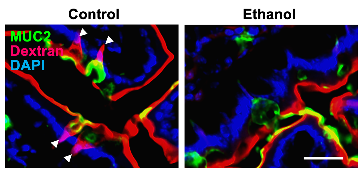
"Scientists have now discovered how alcohol can switch off an immune "alarm system" in the gut, allowing bad bacteria to escape their natural habitat to flood into the liver, rapidly causing inflammation to the organ. This bacterial invasion is a key driver of the inflammation and injury seen in alcohol-associated liver disease (ALD). ..."

"... Now, scientists ... have found that chronic alcohol use impairs the production of a key cellular signaling protein that helps keep gut bacteria within the gut. Without this guardrail in place, bacteria from the gut can more easily migrate to the liver, exacerbating liver damage caused by alcohol. Targeting this mechanism with existing drugs could provide one approach to minimizing the liver damage from alcohol use and reducing the burden of ALD.

Studying a combination of human liver biopsies and mouse models of ALD, the researchers found:
  • Chronic alcohol use reduced the expression of muscarinic acetylcholine receptor M4 (mAChR4), a key cellular communication protein in the gut.
  • Reduced mAChR4 expression hindered the formation of goblet cell-associated antigen passages (GAPs), specialized structures that teach the immune system to promote antimicrobial immunity, thereby preventing harmful bacteria from migrating to the liver. 
  • Restoring mAChR4 function, either by chemically activating mAChR4 or by targeting related signaling pathways allowed GAPs to form and conferred resistance to ALD. 
"

From the abstract:
"Alcohol-use disorder and alcohol-associated liver disease (ALD) are major causes of death and liver transplantation. The gut–liver axis has a crucial yet poorly understood role in ALD pathogenesis, which depends on microbial translocation.
Intestinal goblet cells (GCs) educate the immune system by forming GC-associated antigen passages (GAPs) on activation of muscarinic acetylcholine receptor M4 (mAChR4, also known as M4), enabling sampling of luminal antigens by lamina propria antigen-presenting cells.
Here we show that chronic alcohol use in humans and mice downregulates small intestinal mAChR4 and reduces GAP formation, disrupting antimicrobial immunity. This is reversed on activation of intestinal IL-6 signal transducer (IL6ST, also known as glycoprotein 130; gp130), which restores mAChR4 expression and GAP formation, enabling induction of downstream type-3 innate lymphoid cell-derived IL-22 and antimicrobial REG3 proteins. This blunts translocation of enteric bacteria to the liver, thereby conferring ALD resistance. GAP induction by GC-specific mAChR4 activation was essential and sufficient for prevention of ethanol-induced steatohepatitis. These results lay the foundation for a therapeutic approach using mAChR4 or IL6ST agonists to promote GAP formation and prevent ALD by inhibiting microbial translocation."

Alcohol's impact on gut immunity and liver health


Alcohol Blocks Gut Immune Gatekeepers, Compromising Defense Against Bacteria in ALD "mAChR4 -regulated goblet cell–associated antigen passages (GAPs) train gut immune cells, acting as sentinels, maintaining antimicrobial defense in the gut-liver axis, and positioning mAChR4 agonism and GAP formation as a therapeutic checkpoint for alcohol-associated liver disease (ALD)."


Alcohol reduces mAChR4 and GAP formation. Figure from Llorente et al., Nature, 2025


Tuesday, May 20, 2025

Key gene found to control liver's choice between sugar and fat storage

Amazing stuff!

"The research team found that when the PPP1R3B gene is more active, the liver tends to store more energy as glycogen. The liver stores more energy as fat when the gene is less active. This shift between glycogen and fat storage is crucial because it affects how the body manages blood sugar and fat levels. ..."

From the abstract:
"The PPP1R3B gene, encoding PPP1R3B protein, is critical for liver glycogen synthesis and maintaining blood glucose levels. Genetic variants affecting PPP1R3B expression are associated with several metabolic traits and liver disease, but the precise mechanisms are not fully understood.
We studied the effects of both Ppp1r3b overexpression and deletion in mice and cell models and found that both changes in Ppp1r3b expression result in dysregulated metabolism and liver damage, with overexpression increasing liver glycogen stores, while deletion resulted in higher liver lipid accumulation.
These patterns were confirmed in humans where variants increasing PPP1R3B expression had lower liver fat and decreased plasma lipids, whereas putative loss-of-function variants were associated with increased liver fat and elevated plasma lipids. These findings support that PPP1R3B is a crucial regulator of hepatic metabolism beyond glycogen synthesis and that genetic variants affecting PPP1R3B expression levels influence if hepatic energy is stored as glycogen or triglycerides."

Key gene found to control liver's choice between sugar and fat storage

Scientists Discover Key Gene Impacts Liver Energy Storage, Affecting Metabolic Disease Risk (original news release) "A new study ... reveals that a single gene plays a big role in how the liver stores energy, a process that’s critical for overall health and for managing diseases like type 2 diabetes. ... the research focuses on the PPP1R3B gene. This gene tells the liver how to handle energy: store it as glycogen (a form of sugar) or triglycerides (a type of fat)."


Fig. 1. Mice with hepatocyte Ppp1r3b deletion develop metabolic dysfunction contributing to hepatic steatosis with age, while Ppp1r3b-overexpressing mice rapidly increase glycogen synthesis and storage contributing to liver damage with aging.


Sunday, April 14, 2024

LyGenesis Announces First Patient Dosed in its Clinical Trial of a First-in-Class Regenerative Cell Therapy for Patients with End-Stage Liver Disease

Good news!

""In a medical first, we have now dosed our first patient in a clinical trial using their own lymph nodes as living bioreactors to regenerate an ectopic organ," ... "This therapy will potentially be a remarkable regenerative medicine milestone by helping patients with ESLD grow new functional ectopic livers in their own body. If our study is successful and we obtain FDA approval, our allogenic cell therapy could enable one donated liver to treat many dozens of ESLD patients, which could help to tilt the current organ supply-demand imbalance in favor of patients." ..."

LyGenesis Announces First Patient Dosed in its Phase 2a Clinical Trial of a First-in-Class Regenerative Cell Therapy for Patients with End-Stage Liver Disease | Lygenesis

‘Mini liver’ will grow in person’s own lymph node in bold new trial Biotechnology firm LyGenesis has injected donor cells into a person with liver failure for the first time.


Researchers injected a mouse’s lymph node with liver cells (green), converting the organ into a ‘mini liver’.


Monday, March 25, 2024

Aspirin cuts liver fat in trial

The good old Aspirin (in use for over 2,400 years) keeps on giving! I look forward to take my daily baby (low dose) Aspirin later today! 😊 Long live the liver!

"... In their Phase 2 trial, 80 adults with MASLD were randomized to receive daily low-dose aspirin or placebo for six months. At the end of the study, the average change in liver fat content was minus 6.6 percent with aspirin versus plus 3.6 percent with placebo, showing that low-dose aspirin reduced the average liver fat content by 10.2 percent compared with placebo. The aspirin was found to be safe and well-tolerated, and also improved various markers of liver health. ..."

From the key points and abstract:
"Key Points
Question
In patients with metabolic dysfunction–associated steatotic liver disease (MASLD), does 81 mg of aspirin daily reduce the quantity of hepatic fat at 6-month follow-up compared with placebo?
Findings
In this phase 2 randomized clinical trial of 80 individuals with MASLD, daily aspirin reduced the quantity of hepatic fat at 6-month follow-up compared with placebo (mean difference, −10.2%).
Meaning
In a preliminary randomized clinical trial of adults with MASLD, 6 months of daily low-dose aspirin significantly reduced liver fat content compared with placebo, but findings are preliminary and require confirmation in a larger population.
Abstract
Importance
Aspirin may reduce severity of metabolic dysfunction–associated steatotic liver disease (MASLD) and lower the incidence of end-stage liver disease and hepatocellular carcinoma, in patients with MASLD. However, the effect of aspirin on MASLD is unknown.
Objective
To test whether low-dose aspirin reduces liver fat content, compared with placebo, in adults with MASLD.
Design, Setting, and Participants
This 6-month, phase 2, randomized, double-blind, placebo-controlled clinical trial was conducted at a single hospital in Boston, Massachusetts. Participants were aged 18 to 70 years with established MASLD without cirrhosis. Enrollment occurred between August 20, 2019, and July 19, 2022, with final follow-up on February 23, 2023.
Interventions
Participants were randomized (1:1) to receive either once-daily aspirin, 81 mg (n = 40) or identical placebo pills (n = 40) for 6 months.
Main Outcomes and Measures
The primary end point was mean absolute change in hepatic fat content, measured by proton magnetic resonance spectroscopy (MRS) at 6-month follow-up. The 4 key secondary outcomes included mean percentage change in hepatic fat content by MRS, the proportion achieving at least 30% reduction in hepatic fat, and the mean absolute and relative reductions in hepatic fat content, measured by magnetic resonance imaging proton density fat fraction (MRI-PDFF). Analyses adjusted for the baseline value of the corresponding outcome. Minimal clinically important differences for study outcomes were not prespecified.
Results
Among 80 randomized participants (mean age, 48 years; 44 [55%] women; mean hepatic fat content, 35% [indicating moderate steatosis]), 71 (89%) completed 6-month follow-up. The mean absolute change in hepatic fat content by MRS was −6.6% with aspirin vs 3.6% with placebo (difference, −10.2% [95% CI, −27.7% to −2.6%]; P = .009). Compared with placebo, aspirin treatment significantly reduced relative hepatic fat content (−8.8 vs 30.0 percentage points; mean difference, −38.8 percentage points [95% CI, −66.7 to −10.8]; P = .007), increased the proportion of patients with 30% or greater relative reduction in hepatic fat (42.5% vs 12.5%; mean difference, 30.0% [95% CI, 11.6% to 48.4%]; P = .006), reduced absolute hepatic fat content by MRI-PDFF (−2.7% vs 0.9%; mean difference, −3.7% [95% CI, −6.1% to −1.2%]; P = .004]), and reduced relative hepatic fat content by MRI-PDFF (−11.7 vs 15.7 percentage points; mean difference, −27.3 percentage points [95% CI, −45.2 to −9.4]; P = .003). Thirteen participants (32.5%) in each group experienced an adverse event, most commonly upper respiratory tract infections (10.0% in each group) or arthralgias (5.0% for aspirin vs 7.5% for placebo). One participant randomized to aspirin (2.5%) experienced drug-related heartburn.
Conclusions and Relevance
In this preliminary randomized clinical trial of patients with MASLD, 6 months of daily low-dose aspirin significantly reduced hepatic fat quantity compared with placebo. Further study in a larger sample size is necessary to confirm these findings."

Aspirin cuts liver fat in trial — Harvard Gazette 10 percent reduction seen in small study of disease that affects up to a third of U.S. adults


Sunday, October 15, 2023

Egypt becomes the first country to achieve WHO validation on the path to elimination of hepatitis C

Good news!

"The World Health Organization (WHO) congratulates Egypt for its unprecedented progress towards eliminating hepatitis C, becoming the first country to achieve “gold tier” status on the path to elimination of hepatitis C as per WHO criteria. Achieving the gold tier means that Egypt has fulfilled the programmatic requirements that facilitate the reduction of new hepatitis C infections and deaths to levels that position the country to end the hepatitis C epidemic.

Globally, 58 million people are living with chronic hepatitis C infection. While there is no vaccine, the disease can be cured with highly effective and curative short-course treatments that last 8–12 weeks. Globally, however, 4 out of 5 people living with hepatitis C do not know that they are infected. Unless treated or cured, the infection can cause liver disease and cancer. ..."

WHO EMRO | Egypt becomes the first country to achieve WHO validation on the path to elimination of hepatitis C | News | Media centre

Wednesday, July 19, 2023

First successful robotic liver transplant performed in the US

Good news! Even surgeons may go unemployed as robotics progresses!

"... The man in his 60s, who required a critical transplant due to liver cancer and cirrhosis caused by hepatitis C, underwent surgery performed by the advanced machinery while the head surgeon controlled the robot’s movements from a console nearby. ..."

First successful robotic liver transplant performed in the US


Friday, May 26, 2023

Cancers in distant organs alter liver function

Amazing stuff! Cancer is history (soon)! The more we know about cancer, the better! Another example how nasty cancer can be!

"Cancers often release molecules into the bloodstream that pathologically alter the liver, shifting it to an inflammatory state, causing fat buildup and impairing its normal detoxifying functions ...
In the study ... the researchers found that a wide variety of tumor types growing outside the liver remotely reprogram the liver to a state resembling fatty liver disease via secretion of extracellular vesicles and particles (EVPs) containing fatty acids. ...
The team also observed that reprogrammed livers have high levels of inflammation, marked by elevated level of tumor necrosis factor-α (TNF-α), and low levels of drug-metabolizing enzymes called cytochrome P450, which break down potentially toxic molecules, including many drug molecules. The observed reduction in cytochrome P450 levels could explain why cancer patients often become less tolerant of chemotherapy and other drugs as their illness progresses. ..."

From the abstract:
"Cancer alters the function of multiple organs beyond those targeted by metastasis. Here we show that inflammation, fatty liver and dysregulated metabolism are hallmarks of systemically affected livers in mouse models and in patients with extrahepatic metastasis. We identified tumour-derived extracellular vesicles and particles (EVPs) as crucial mediators of cancer-induced hepatic reprogramming, which could be reversed by reducing tumour EVP secretion via depletion of Rab27a. All EVP subpopulations, exosomes and principally exomeres, could dysregulate hepatic function. The fatty acid cargo of tumour EVPs—particularly palmitic acid—induced secretion of tumour necrosis factor (TNF) by Kupffer cells, generating a pro-inflammatory microenvironment, suppressing fatty acid metabolism and oxidative phosphorylation, and promoting fatty liver formation. Notably, Kupffer cell ablation or TNF blockade markedly decreased tumour-induced fatty liver generation. Tumour implantation or pre-treatment with tumour EVPs diminished cytochrome P450 gene expression and attenuated drug metabolism in a TNF-dependent manner. We also observed fatty liver and decreased cytochrome P450 expression at diagnosis in tumour-free livers of patients with pancreatic cancer who later developed extrahepatic metastasis, highlighting the clinical relevance of our findings. Notably, tumour EVP education enhanced side effects of chemotherapy, including bone marrow suppression and cardiotoxicity, suggesting that metabolic reprogramming of the liver by tumour-derived EVPs may limit chemotherapy tolerance in patients with cancer. Our results reveal how tumour-derived EVPs dysregulate hepatic function and their targetable potential, alongside TNF inhibition, for preventing fatty liver formation and enhancing the efficacy of chemotherapy."

Cancers in distant organs alter liver function | Cornell Chronicle

Wednesday, October 19, 2022

Livers can stay alive and functional for over 100 years across multiple donors and recipients

Amazing stuff! I toast to that! (just kidney kidding) 😊

"A research team composed of members from the University of Texas (UT) and TransMedics, Massachusetts reports that a human liver can stay functional for over 100 years. The results are based on a (still small, but growing) subset of livers that have been transplanted once or several times for a cumulative age of over 100 years across their patients. ...
Of the 253,406 livers transplanted between 1990 and 2022, 25 met this age criterion. ... These long-lived livers came from older donors. The average donor age of this subset of organs was much higher than that of donors for livers that did not pass 100 years of age: 84.7 years vs. 38.5 years of age, respectively. Another notable difference between the two groups that the team observed was that donors from the centenarian liver group had lower incidences of diabetes and fewer donor infections. ..."

Livers can stay alive and functional for over 100 years across multiple donors and recipients Such findings pave the way towards older people being eligible to donate their livers and saving more lives.

Sunday, August 07, 2022

Tissue model reveals key players in liver regeneration

Good news!

"The human liver has amazing regeneration capabilities: Even if up to 70 percent of it is removed, the remaining tissue can regrow a full-sized liver within months. ...
The new study, which appears this week in the Proceedings of the National Academy of Sciences, has identified one molecule that appears to play a key role, and also yielded several other candidates that the researchers plan to explore further. ...
One key factor is the reciprocal relationship between hepatocytes (the main type of cell found in the liver) and endothelial cells, which line the blood vessels. Hepatocytes produce factors that help blood vessels develop, and endothelial cells generate growth factors that help hepatocytes proliferate.
Another contributor that researchers have identified is fluid flow in the blood vessels. ...
To model all of these interactions ... designs microfluidic devices with channels that mimic blood vessels. To create these models of “regeneration on a chip,” the researchers grew blood vessels along one of these microfluidic channels and then added multicellular spheroid aggregates derived from liver cells from human organ donors. ...
Using this system, the researchers showed that increased fluid flow on its own did not stimulate hepatocytes to enter the cell division cycle. However, if they also delivered an inflammatory signal (the cytokine IL-1-beta), hepatocytes did enter the cell cycle. ..."

"Liver disease causes ∼2 million annual deaths, yet medical treatments and transplantable organs are both lacking. The liver can regenerate when mature hepatocytes divide, and while this process is well studied in rodents, parallel study of human biology has been impossible. We developed a microfluidic device that allows us to manipulate fluid flow, circulating cytokines, and/or paracrine interactions between liver and vascular cells, in order to model multicellular aspects of human liver regeneration. We found that physiologically relevant shear stresses increased the secretion of angiogenesis- and regeneration-associated factors, including prostaglandin E2 from endothelial cells, and induced primary human hepatocytes to enter the cell cycle. ..."

From the abstract:
"... To this end, we developed a three-dimensional (3D) platform called structurally vascularized hepatic ensembles for analyzing regeneration (SHEAR) to model multiple aspects of human liver regeneration. SHEAR enables control over hemodynamic alterations to mimic those that occur during liver injury and regeneration and supports the administration of biochemical inputs such as cytokines and paracrine interactions with endothelial cells. We found that exposing the endothelium-lined channel to fluid flow led to increased secretion of regeneration-associated factors. Stimulation with relevant cytokines not only amplified the secretory response, but also induced cell-cycle entry of primary human hepatocytes (PHHs) embedded within the device. Further, we identified endothelial-derived mediators that are sufficient to initiate proliferation of PHHs in this context. ..."

Tissue model reveals key players in liver regeneration | MIT News | Massachusetts Institute of Technology


Sunday, July 12, 2020

Boosting a liver protein may mimic the brain benefits of exercise

Good news! Towards a pill/remedy for everything or the fountain of youth! Very soon you may not need a treadmill or jogging anymore (just kidding)! :-)


"Indeed, aged mice that received plasma from young or old mice that had exercised showed beneficial effects in their brains without hitting the treadmill. ... Plasma concentrations of glycosylphosphatidylinositol (GPI)–specific phospholipase D1 (Gpld1), a GPI-degrading enzyme derived from liver, were found to increase after exercise and to correlate with improved cognitive function in aged mice, and concentrations of Gpld1 in blood were increased in active, healthy elderly humans. Increasing systemic concentrations of Gpld1 in aged mice ameliorated age-related regenerative and cognitive impairments by altering signaling cascades downstream of GPI-anchored substrate cleavage. We thus identify a liver-to-brain axis by which blood factors can transfer the benefits of exercise in old age."


Boosting a liver protein may mimic the brain benefits of exercise | Science News Liver-made proteins that circulate in the blood improved memories, a mouse study suggests


Here is a link to the underlying paper:

Blood factors transfer beneficial effects of exercise on neurogenesis and cognition to the aged brain