Showing posts with label neurological disorder. Show all posts

Friday, May 01, 2026

Damage to brain’s white matter may play key role in neurodegenerative disease, and could be target for future treatments

Good news!

"... The team ... created localised damage to myelin – the main component of white matter – in a well-defined brain circuit and followed what happened over time. They found that small, localised myelin damage triggered a striking response in a connected, remote grey matter region. Neuronal activity fell, microglia – the brain’s immune cells – became activated, and connections between neurons were lost.

Crucially, these changes were not permanent. After myelin was regenerated, neuronal activity recovered, connections between neurons returned, and the inflammatory response subsided.

The study also challenges a common assumption about brain inflammation. Grey matter inflammation is traditionally viewed as harmful. But here, the team found that this transient response was part of the repair process itself. When they prevented grey matter inflammation, myelin regeneration was impaired.

Conversely, when the team blocked myelin regeneration, the grey matter response did not resolve and instead became chronic. This suggests that failed myelin regeneration may help drive the persistent low-grade inflammation seen in neurodegenerative disease. ..."

"Focal white matter lesions occur in most neurodegenerative disorders. Despite occurring early in disease, white matter lesions are considered to be independent of, or secondary to, grey matter neuroinflammation, synapse loss and altered neuronal activity. Notably, their functional effect on neuronal circuits remains understudied.
To address this, we generated a focal white matter lesion in the rat brain within a clinically relevant, anatomically well-defined circuit, in which these lesions occur in many neurodegenerative disorders.
Here we show that focal white matter lesions evoke transient neuronal activity changes and microgliosis, with subsequent synapse loss and increased microglial engulfment in the grey matter, which is reversed if myelin regeneration completes. Grey matter microgliosis is often considered to be detrimental; however, we show that it is an integral part of regeneration and is conserved across three distinct mouse circuits and lesioning methods.
Preventing these transient changes in the grey matter blocks myelin regeneration in the white matter.
Conversely, inducing myelin regeneration failure leads to chronic grey matter neuroinflammation. This recapitulates the low-grade inflammation considered to be a dominant mechanism underlying neurodegeneration.
Our findings reveal a form of regenerative plasticity coupling white matter integrity to grey matter function, which may underlie multiple neurodegenerative conditions, and highlight the potential of targeting myelin regeneration to prevent chronic neuroinflammation."

Damage to brain’s white matter may play key role in neurodegenerative disease, and could be target for future treatments | University of Cambridge "Damage to white matter in the brain can trigger features associated with neurodegenerative disease ..."


Fig. 3: Focal white matter lesions evoke changes in microglial states and neuronal mitochondria.


Saturday, February 21, 2026

Reprogramming of brain glial cells to corticospinal neurons may treat ALS and spinal cord injuries

Good news!

"Harvard stem cell biologists have discovered a way to grow the type of brain cells that degenerate in patients with amyotrophic lateral sclerosis (ALS) and suffer damage in spinal cord injuries.

In a paper published in the journal eLife, researchers engineered a cocktail of molecular signals to coax some “progenitor cells” — precursors that can differentiate into other cell types — to generate corticospinal neurons (CSNs), brain cells vital to voluntary motor control. ...

“.progenitor population is that it’s already distributed throughout the brain ... They’re sitting there — resident stem cells.”

The new study offers the first-ever model for growing corticospinal neurons in the lab, opening new windows for researching and potentially regenerating neurons for two devastating neurological afflictions. ..."

"eLife Assessment
This study presents fundamental new findings introducing a new approach for the reprogramming of brain glial cells to corticospinal neurons. The data is highly compelling, with multiple lines of evidence demonstrating the success of this new assay. These exciting findings set the stage for future studies of the potential of these reprogrammed cells to form functional connections in vivo and their utility in clinical conditions where corticospinal neurons are compromised."

From the abstract:
"Corticospinal neurons (CSN) centrally degenerate in amyotrophic lateral sclerosis (ALS), along with spinal motor neurons, and loss of voluntary motor function in spinal cord injury (SCI) results from damage to CSN axons.
For functional regeneration of specifically affected neuronal circuitry in vivo, or for optimally informative disease modeling and/or therapeutic screening in vitro, it is important to reproduce the type or subtype of neurons involved. No such appropriate in vitro models exist with which to investigate CSN selective vulnerability and degeneration in ALS, or to investigate routes to regeneration of CSN circuitry for ALS or SCI, critically limiting the relevance of much research. Here, we identify that the HMG-domain transcription factor Sox6 is expressed by a subset of NG2+ endogenous cortical progenitors in postnatal and adult cortex, and that Sox6 suppresses a latent neurogenic program by repressing proneural Neurog2 expression by progenitors.
We FACS-purify these progenitors from postnatal mouse cortex and establish a culture system to investigate their potential for directed differentiation into CSN. We then employ a multi-component construct with complementary and differentiation-sharpening transcriptional controls (activating Neurog2, Fezf2, while antagonizing Olig2 with VP16:Olig2).
We generate corticospinal-like neurons from SOX6+/NG2+ cortical progenitors and find that these neurons differentiate with remarkable fidelity compared with corticospinal neurons in vivo. They possess appropriate morphological, molecular, transcriptomic, and electrophysiological characteristics, without characteristics of the alternate intracortical or other neuronal subtypes. We identify that these critical specifics of differentiation are not reproduced by commonly employed Neurog2-driven differentiation. Neurons induced by Neurog2 instead exhibit aberrant multi-axon morphology and express molecular hallmarks of alternate cortical projection subtypes, often in mixed form. Together, this developmentally-based directed differentiation from cortical progenitors sets a precedent and foundation for in vitro mechanistic and therapeutic disease modeling, and toward regenerative neuronal repopulation and circuit repair."

A ‘cocktail’ recipe for brain cells — Harvard Gazette "Stem cell biologists discover how to regenerate type damaged in ALS, spinal cord injuries"


Fig. 1 Identification and culture of SOX6+/NG2+ cortical progenitors with high purity and fidelity


Tuesday, February 17, 2026

Neue MRT-Analyse deckt Fehldiagnosen bei Parkinson auf

Good news! Empfehlenswert!

"Parkinson – oder doch etwas anderes? Diese Frage beschäftigt ... Neurologen seit Jahren. Denn nicht jede Bewegungsstörung mit Zittern, Steifheit und Stürzen ist Morbus Parkinson. Zwei seltene, aber relevante Erkrankungen werden häufig übersehen: die progressive supranukleäre Lähmung (PSP) und die kortikobasale Degeneration (CBD).

Eine internationale Studie ... dass moderne Magnetresonanztomographie (MRT) hier deutlich mehr Klarheit schafft. Die Ergebnisse wurden im Journal of Prevention of Alzheimer’s Disease veröffentlicht. ...

Bei PSP zeigten sich vor allem Veränderungen im Hirnstamm und in tiefen Kerngebieten. Bestimmte kortikale Regionen waren selektiv mitbetroffen.
CBD hingegen wies stärker ausgeprägte Schäden in der Großhirnrinde auf, insbesondere in Arealen für Motorik und sensorische Integration.

„Obwohl sie klinisch sehr ähnlich aussehen mögen, schädigen PSP und CBD das Gehirn auf unterschiedliche Weise“, ... „Diese Unterschiede spiegeln sich im MRT wider, und indem wir sie zu einem Signaturmuster kombinieren, können wir viel besser bestimmen, an welcher Krankheit der jeweilige Patient leidet.“ ..."

Neue MRT-Analyse deckt Fehldiagnosen bei Parkinson auf "Ein neues MRT-Analyseverfahren unterscheidet PSP und CBD zuverlässig von Parkinson – und macht klinische Studien deutlich effizienter."

Thursday, February 12, 2026

FDA Approves tDCS headset for Depression Treatment at home

Good news!

"For years, a small group of technology enthusiasts have been applying gentle electrical current to their brains in an effort to gain cognitive benefits, improve sleep, or aid memory. While brain stimulation, also referred to as neuromodulation, can take many forms, transcranial direct current stimulation (tDCS) emerged as a reasonably safe, affordable choice for at-home experimentation for a range of purposes.

These devices have often been home-brewed or sold as wellness tools, but in her research into the do-it-yourself tDCS community, Anna Wexler, a medical ethicist at the University of Pennsylvania, found that in addition to brain boosting, many practitioners were self-medicating, using electrotherapy to treat symptoms of depression and anxiety. Until recently, there were no medical tDCS devices with U.S. Food and Drug Administration approval.

In December the FDA approved a tDCS headset produced by Flow Neuroscience for treatment of major depressive disorder. ..."

FDA Approves tDCS for Depression Treatment - IEEE Spectrum "Flow’s headset is the first tDCS device approved by the FDA"


Flow Neuroscience’s transcranial direct current stimulation device has been approved by the FDA to treat depression


Friday, February 06, 2026

Multiple sclerosis may have two distinct biological pathways

Good news!

"... For decades, multiple sclerosis (MS) has been defined primarily by its symptoms, rather than its underlying biology. Now, a new study aims to challenge that approach, presenting evidence that MS may actually follow two distinct biological pathways. ... 

To do that, the team paired two complementary sources of information. One came from blood measurements of serum neurofilament light chain (sNfL), a protein released when nerve cells are damaged and widely used as a marker of disease activity. The other came from MRI scans that captured how structural degeneration spread through the brain over time.

Rather than examining each dataset in isolation, the researchers analyzed them together using a machine-learning system developed at UCL called SuStaIn (Subtype and Stage Inference). The model is designed to detect subtle disease patterns and map how they evolve, allowing the team to test whether MS follows a single biological trajectory or something more complex. ...

Instead of detecting a smooth disease spectrum, two distinct structural patterns emerged. The team found that patients clustered into separate groups that reflected different underlying pathways of neurodegeneration.

One subtype was marked by early damage concentrated in the brain’s cortex, while the other was dominated by degeneration in white matter regions. Although both patterns ultimately produced the symptoms associated with multiple sclerosis, the location of tissue damage and the path it followed through the brain differed substantially between the two groups. ..."

From the abstract:
"Multiple sclerosis (MS) is a highly heterogeneous disease in its clinical manifestation and progression. Predicting individual disease courses is key for aligning treatments with underlying pathobiology.
We developed an unsupervised machine learning model integrating MRI-derived measures with serum neurofilament light chain (sNfL) levels to identify biologically informed MS subtypes and stages. ...

In comparison to MRI-only models, incorporating sNfL with MRI improved correlations of data-derived stages with the Expanded Disability Status Scale in the training (Spearman’s ρ = 0.420 versus MRI-only ρ = 0.231, P = 0.001) and external test sets (ρ = 0.163 for MRI–sNfL, versus ρ = 0.067 for MRI-only).
The early-sNfL subtype showed elevated sNfL, corpus callosum injury and early lesion accrual, reflecting more active inflammation and neurodegeneration, whereas the late-sNfL group showed early volume loss in the cortical and deep grey matter volumes, with later sNfL elevation.
Cross-sectional subtyping predicted longitudinal radiological activity: the early-sNfL group showed a 144% increased risk of new lesion formation (hazard ratio = 2.44, 95% confidence interval 1.38–4.30, P < 0.005) compared with the late-sNfL group. Baseline subtyping, over time, predicted treatment effect on new lesion formation on the external test set (faster lesion accrual in early-sNfL compared with late-sNfL, P = 0.01), in addition to treatment effects on brain atrophy (early sNfL average percentage brain volume change: −0.41, late-sNfL = −0.31, P = 0.04).

Integration of sNfL provides an improved framework in comparison to MRI-only subtyping of MS to stage disease progression and inform prognosis. Our model predicted treatment responsiveness in early, more active disease states. This approach offers a powerful alternative to conventional clinical phenotypes and supports future efforts to refine prognostication and guide personalized therapy in MS."

Multiple sclerosis may have two distinct biological pathways


Fig. 1 Overview of the study


Monday, January 26, 2026

New brain atlas of lysosomal proteins could help researchers studying neurological disease

Good news!

"In brief
  • Stanford researchers created the first atlas of lysosomal proteins in the brain, crucial for understanding neurodegenerative diseases.
  • The atlas highlights links between lysosomal dysfunction and disorders like Alzheimer’s and Parkinson’s, guiding potential therapeutic developments.
  • The data is publicly accessible, promoting global collaboration and exploration of lysosomal roles in neurological conditions.
...

“Lysosomal function is essential for brain health, and mutations in lysosomal genes are risk factors for neurodegenerative diseases,” ..."

From the highlights and abstract:
"Highlights
• Lysosomal proteomics of brain cell types
• Identification of previously uncharacterized and cell-type-specific lysosomal proteins
• SLC45A1 is a neuron-specific lysosomal sugar transporter
• SLC45A1 loss drives lysosomal and mitochondrial dysfunction

Summary
Mutations in lysosomal genes cause neurodegeneration and neuronopathic lysosomal storage disorders (LSDs). Despite their essential role in brain homeostasis, the cell-type-specific composition and function of lysosomes remain poorly understood.
Here, we report a quantitative protein atlas of lysosomes from mouse neurons, astrocytes, oligodendrocytes, and microglia. We identify dozens of proteins not previously annotated as lysosomal and reveal the diversity of lysosomal composition across brain cell types.
Notably, we identified SLC45A1, a gene whose mutations cause a monogenic neurological disease, as a neuron-specific lysosomal protein.
Loss of SLC45A1 causes lysosomal dysfunction in vitro and in vivo. SLC45A1 functions as a lysosomal sugar transporter and impacts the stability of the V1 subunits of the vacuolar ATPase (V-ATPase). Consistently, SLC45A1 loss reduces lysosomal V1 subunits, elevates lysosomal pH, and disrupts iron homeostasis, causing mitochondrial dysfunction. Altogether, our work redefines SLC45A1-associated disease as an LSD and establishes a comprehensive map to study lysosome biology at cell-type resolution."

New atlas could help researchers studying neurological disease | Stanford Report "A database of lysosomal proteins is already guiding researchers in studying how brain cells’ waste and recycling systems work – or don’t – in Alzheimer’s and other neurological diseases."


Graphical abstract


Thursday, January 22, 2026

Bavisant identified as a therapeutic candidate for multiple sclerosis via drug repurposing

Good news!

From the editor's summary and abstract:
"Editor’s summary
Drug repurposing could allow fast and cost-effective identification of neuroprotective and remyelination therapies for multiple sclerosis. Gacem et al. performed in silico screening to find potential candidates in a library of 1500 repurposed drugs. These drug candidates were tested in cell models to identify compounds with low toxicity profiles and potential for remyelination. The therapeutic potential of one lead compound, the histamine receptor H3 antagonist bavisant, was validated by proof-of-principle experiments in several mouse models of demyelination. These findings suggest that the presented screening pipeline could be valuable to identify additional repurposed compounds with potential remyelinating and neuroprotective properties. ...

Abstract
Current treatments for multiple sclerosis (MS) are insufficient to delay the neurodegenerative process that is the main cause of disability progression in patients with MS. Therapeutics aimed at supporting myelin regeneration and neuroprotection are thus a major unmet medical need for the progressive forms of MS.
To address this, we developed a strategy combining in silico screening of more than 1500 repurposed compounds with a validation pipeline of models, encompassing rodent and human in vitro assays as well as mouse models of demyelination/remyelination.
From the initial library, 273 drugs were prioritized in silico on the basis of the predicted effects on myelination and neuroprotection, and among them, 160 were potentially nontoxic.
We identified 32 molecules that exerted a promyelinating and a neuroprotective action on rodent and human oligodendroglia and neurons. Our data identified classes of compounds with potentially distinct mechanisms of action that may foster remyelination and neuroprotection.
The therapeutic activity of one selected drug, the histamine receptor H3 antagonist bavisant, was further validated in mouse models of demyelination and axonal injury reproducing some key pathological features occurring in MS.
Our in vivo studies demonstrated that bavisant promoted remyelination and neuroprotection when administered to LPC-treated, cuprizone-fed, or MOG-induced EAE mice, as well as in a human oligodendroglia chimeric mouse model of demyelination/remyelination.
These findings provide proof-of-concept validation for bavisant as a candidate for neuroprotective clinical trials in MS."

In silico screening and preclinical validation identify bavisant as a therapeutic candidate for multiple sclerosis | Science Translational Medicine (no public access)

Wednesday, January 07, 2026

Hörgeräte schützen vor Demenz

Ich wette, da ist was dran, dass verschlechtertes Hören im Alter u. U. zu Demenz führen oder beitragen kann.

Manche ältere Menschen realisieren vielleicht nicht einmal wie weit ihre Hörfähigkeit über die Jahre nachgelassen hat.

Nicht gut hören macht auch einsam!

"Schamgefühle gehören neben Praktikabilitätsgründen zu den häufigsten Gründen, warum Menschen mit Schwerhörigkeit Hörhilfen ablehnen. Hörgeräte werden noch immer mit Schwäche und Altwerden assoziiert, und viele befürchten negative Reaktionen im sozialen Umfeld. Dazu kommt, dass viele Betroffene ihr eigenes Hördefizit lange unterschätzen, was eine Korrektur durch Hörgeräte und eine schnelle Eingewöhnung verzögert – mit dem Ergebnis, dass bis ins hohe Alter keine Hörhilfen getragen werden. Doch Schwerhörigkeit ist kein Komfort-Problem, sie birgt eine ­Gefahr: Denn wer nicht gut hört, hat ein nachweisbar erhöhtes Demenzrisiko. ..."

Hörgeräte schützen vor Demenz | FAZ (erfordert Abo) "Bis zu sieben Prozent aller Demenzerkrankungen könnten verhindert werden, wenn Menschen besser auf ihr Gehör achtgeben würden. Das könnte auch Einsamkeit verhindern."

Tuesday, December 30, 2025

Alzheimer's disease can be reversed in animal models to achieve full neurological recovery

Good news! This could be a major breakthrough!

"... Through studying diverse preclinical mouse models and human AD brains, the team showed that the brain's failure to maintain normal levels of a central cellular energy molecule, NAD+, is a major driver of AD, and that maintaining proper NAD+ balance can prevent and even reverse the disease.

NAD+ levels decline naturally across the body, including the brain, as people age. Without proper NAD+ balance, cells eventually become unable to execute critical processes required for proper functioning and survival.

In this study, the team showed that the decline in NAD+ is even more severe in the brains of people with AD, and that this also occurs in mouse models of the disease. ...

the research team tested whether preventing the loss of brain NAD+ balance before disease onset, or restoring brain NAD+ balance after significant disease progression, could prevent or reverse AD, respectively.

The study was based on their previous work, published in Proceedings of the National Academy of Sciences, showing that restoring the brain's NAD+ balance achieved pathological and functional recovery after severe, long-lasting traumatic brain injury. They restored NAD+ balance by administering a now well-characterized pharmacological agent known as P7C3-A20, developed in the Pieper lab.

Remarkably, not only did preserving NAD+ balance protect mice from developing AD, but delayed treatment in mice with advanced disease also enabled the brain to fix the major pathological events caused by the genetic mutations. Moreover, both lines of mice fully recovered cognitive function.

This was accompanied by normalized blood levels of phosphorylated tau 217, a recently approved clinical biomarker of AD in people ..."

From the highlights and abstract:
"Highlights
• Severity of Alzheimer’s disease (AD) correlates with NAD+ homeostasis dysregulation
• Preserving brain NAD+ homeostasis prevents AD in mice
• Restoring brain NAD+ homeostasis reverses advanced AD in mice
• Multiomics across human and mouse AD brain identifies nodes for human AD reversal

Summary
Alzheimer’s disease (AD) is traditionally considered irreversible.
Here, however, we provide proof of principle for therapeutic reversibility of advanced AD. In advanced disease amyloid-driven 5xFAD mice, treatment with P7C3-A20, which restores nicotinamide adenine dinucleotide (NAD+) homeostasis, reverses tau phosphorylation, blood-brain barrier deterioration, oxidative stress, DNA damage, and neuroinflammation and enhances hippocampal neurogenesis and synaptic plasticity, resulting in full cognitive recovery and reduction of plasma levels of the clinical AD biomarker p-tau217.
P7C3-A20 also reverses advanced disease in tau-driven PS19 mice and protects human brain microvascular endothelial cells from oxidative stress.
In humans and mice, pathology severity correlates with disruption of brain NAD+ homeostasis, and the brains of nondemented people with Alzheimer’s neuropathology exhibit gene expression patterns suggestive of preserved NAD+ homeostasis.
Forty-six proteins aberrantly expressed in advanced 5xFAD mouse brain and normalized by P7C3-A20 show similar alterations in human AD brain, revealing targets with potential for optimizing translation to patient care."

Alzheimer's disease can be reversed in animal models to achieve full neurological recovery

New study shows Alzheimer’s disease can be reversed to achieve full neurological recovery—not just prevented or slowed—in animal models (original news release) "Researchers from Case Western Reserve University, University Hospitals and the Cleveland VA showed restoring brain’s energy balance led to both pathological and functional recovery"


Graphical abstract


Figure 1 P7C3-A20 restores brain NAD+ homeostasis, cognitive function, and synaptic plasticity in aged symptomatic 5xFAD mice


Tuesday, December 09, 2025

Overcoming dementia/Alzheimers with digital twins

Would it not be great for an individual with onset of dementia to record daily activities and to commit important documents, events etc. to memory of the digital twin? I bet this can be realized in the very near future.

The following article is not directly about this subject, but about biological digital twins for diagnosis and treatment. However, the article gave me the idea!

Your digital twin might save your life — Harvard Gazette

Just discovered that PNAS also just published an article about digital twins: Digital twins come to the life sciences

Wednesday, November 05, 2025

Long-term study challenges assumptions about epilepsy recovery

Good news!

"Patients with treatment-resistant epilepsy often cycle through multiple medications as they seek relief from the seizures that disrupt their lives. Yet in many cases, these drugs offer little benefit, reinforcing the long-held belief among experts that treatment-resistant epilepsy is a condition that remains stable at best — or gradually worsens over time.

A new study, however, challenges this longstanding notion by showing that a subset of these hard-to-treat patients do experience seizure relief, though researchers aren’t exactly sure what’s driving the improvement. ...

“The conventional wisdom is that once you’ve failed two medications, the likelihood of the third medication, or the fourth, making a patient seizure-free is less than 5%,” ...

“But we showed that there are some people who do improve somewhat over time, even after three, four, or five medications, which was surprising. 

“There was even a small group — about 17% of people — who were seizure-free for three months, which is a big deal because it challenges the current understanding and shows that people can improve,” ...

Were new or ongoing treatments responsible for the improvement, or does seizure frequency simply wane over time? Researchers are still analyzing the data. ...

Globally, the prevalence of epilepsy ranges between 0.6%-1.4% across respective populations, ... The good news is that many people — 40% to 60% of patients — become seizure-free when they begin anti-seizure medication, research shows. Another 30% have the medically refractory type that doesn’t respond to medication. ..."

From the abstract:
"Importance:
Open-label trials of antiseizure medications (ASMs) and devices suggest seizure reduction in focal treatment-resistant epilepsy (FTRE) may demonstrate treatment-related disease-modifying effects. Understanding FTRE trends can provide insight into treatment responses.

Objective:
To determine whether seizure frequency in FTRE improves over time.

Design, setting, and participants:
The Human Epilepsy Project 2 was a prospective, observational, multicenter study of patients with FTRE from May 2018 to September 2021 who were followed up for 18 to 36 months at 10 US-based comprehensive epilepsy centers. Analysis was performed from 2021 to 2024. Study data included seizure frequency, medication use, device use, surgeries tracked using daily electronic diaries, monthly check-ins, medical record review, and case report forms. Eligibility criteria included focal epilepsy diagnosis, age between 16 and 65 years, and failure of 4 or more ASMs (≥2 due to seizure control failure). Participants were recruited as a volunteer sample.

Exposures:
Participants were treated with multiple interventions at their physicians' discretion.

Main outcomes and measures:
The primary outcome was seizure frequency trends, evaluated by quantifying seizure freedom rates and frequency reductions. Medication and device treatment responses were assessed by tracking ASM and device changes.

Results:
Of 196 approached participants, 146 met eligibility criteria and were included in the study. Mean (SD) participant age was 40 (12) years, and epilepsy was diagnosed at a mean (SD) age of 19.8 (13.6) years. The cohort had 84 (57.5%) female participants. A total of 35 participants had implantable devices; 1 had epilepsy surgery during the study. Of 146 participants, 128 provided sufficient seizure data for analysis, and 2 were excluded as outliers. Seizure frequency was reduced in 86 participants (68.3%) during the second half of study participation compared to the first half. In the overall cohort, mean modeled monthly seizure frequency percentage reduction was 68.73% (95% CI, 52.92%-84.54%). From 0 to 12 months (cohort 1), mean modeled percentage reduction was 67.76% (95% CI, 19.42%-116.09%); for 12 to 24 months (cohort 2), 36.00% (95% CI, 9.27%-53.46%); and for longer than 24 months (cohort 3), 66.03% (95% CI, 48.25%-83.80%) (all P < .001).
An ASM was added in 69 participants (54.7%), of whom 46 (66.7%) experienced seizure frequency reduction, including seizure freedom. Seizure trajectories in participants with devices did not significantly differ from those without devices.

Conclusions and relevance:
Findings from the HEP2 study imply that FTRE improves over time, ASM additions had low probability of achieving seizure freedom but contributed to seizure reduction, and device-treated participants exhibited similar seizure trajectories to those without devices. Whether improvements reflected the natural history of FTRE or active management remains unclear, but our findings suggest cautious interpretation of open-label studies positing disease-modifying effects and further research into FTRE treatment response."

Long-term study challenges assumptions about epilepsy recovery | Yale News "Collaborative, long-term study reveals that patients with treatment-resistant epilepsy can improve over time with, and sometimes without, any treatment."

Tuesday, November 04, 2025

Just 5,000 steps a day could slow cognitive decline in people with signs of Alzheimer’s, study suggests. Really!

That is a lot of steps per day! I think, this recommendation might be excessive! What about other physical exercises/activities?

What is the recommendation now? 5,000, 8,000 or 10,000 steps per day? Maybe next year we are told 3,000 steps per day! 😊

It appears, the recommendations are all over the place! A sedentary individual may just lean back pondering all these various recommendations! Caution: irony.

Indeed, the article says the beneficial effect "plateaus at around 5,000 to 7,500 steps per day".

Google Fit tells me that my typical about 3 mile walk (not daily) is roughly 6000 steps.

"... The scientists found that patients who started with high levels of beta-amyloid, an early biological sign of Alzheimer’s, declined less if they were more physically active. Low or moderate levels of physical activity in this group, the authors reported, could slow cognitive decline by half compared with inactive individuals. That effect plateaued at around 5,000 to 7,500 steps a day. ..."

"... The research indicates that people don’t need to take 10,000 steps a day, a goal that is often touted but might be hard for some older individuals to attain ..."

"... Cognitive decline was delayed by three years on average for people who walked just 3,000-5,000 steps per day, and by seven years in people who walked 5,000-7,500 steps per day. Sedentary individuals had a significantly faster buildup of tau proteins in the brain and more rapid declines in cognition and daily functioning. ..."

From the abstract:
"Physical inactivity is a recognized modifiable risk factor for Alzheimer’s disease (AD), yet its relationship with progression of AD pathology in humans remains unclear, limiting the effective translation into prevention trials.
Using pedometer-measured step counts in cognitively unimpaired older adults, we demonstrated an association between higher physical activity and slower cognitive and functional decline in individuals with elevated baseline amyloid. Importantly, this beneficial association was not related to lower amyloid burden at baseline or longitudinally.
Instead, higher physical activity was associated with slower amyloid-related inferior temporal tau accumulation, which significantly mediated the association with slower cognitive decline.
Dose–response analyses further revealed a curvilinear relationship, where the associations with slower tau accumulation and cognitive decline reached a plateau at a moderate level of physical activity (5,001–7,500 steps per day), potentially offering a more approachable goal for older sedentary individuals [???].
Collectively, our findings support targeting physical inactivity as an intervention to modify the trajectory of preclinical AD in future prevention trials, and further suggest that preferentially enrolling sedentary individuals with elevated amyloid may maximize the likelihood of demonstrating a protective effect of physical activity on tau accumulation and cognitive and functional decline in early AD."

Take steps to slow Alzheimer's progression: 5,000 of them, actually | STAT "Exercise linked to slower buildup of tau protein in the brain"

Alzheimer’s decline slows with just a few thousand steps a day "A modest increase in physical activity can delay cognitive decline by three years — or more."


Longer walks beat shorter strolls for heart health (no public access) "People who rack up most of their daily steps in walks lasting less than five minutes have a higher risk of cardiovascular disease than do those who amass their steps in big blocks."


Even people who don’t reach the recommended 8,000 steps per day can reap health benefits if they take relatively long walks.


Extended Data Fig. 2: Interactive association between baseline physical activity and Aβ burden on initial ITC tau burden.


Sunday, November 02, 2025

Oral bacteria metabolites linked to Parkinson's via the gut-brain axis

Good news!

"... Korean researchers have uncovered compelling evidence that oral bacteria, once colonized in the gut, can affect neurons in the brain and potentially trigger Parkinson's disease. ...

They have identified the mechanism by which metabolites produced by oral bacteria in the gut may trigger the development of Parkinson's disease. ...

Although previous studies suggested that the gut microbiota of individuals with Parkinson's differs from that of healthy individuals, the specific microbes and metabolites have remained unclear.

They found an increased abundance of Streptococcus mutans—a well-known oral bacterium that causes dental caries—in the gut microbiome of Parkinson's patients.

More importantly, S. mutans produces the enzyme urocanate reductase (UrdA) and its metabolite imidazole propionate (ImP), both of which were present at elevated levels in the gut and blood of patients. ImP appeared capable of entering systemic circulation, reaching the brain, and contributing to the loss of dopaminergic neurons.

Using mouse models, the researchers introduced S. mutans into the gut or engineered E. coli to express UrdA.

As a result, the mice showed elevated ImP levels in blood and brain tissue, along with the hallmark features of Parkinson's symptoms: loss of dopaminergic neurons, heightened neuroinflammation, impaired motor function, and increased aggregation of alpha-synuclein, a protein central to disease progression.

Further experiments demonstrated that these effects depend on the activation of the signaling protein complex mTORC1. Treating mice with an mTORC1 inhibitor significantly reduced neuroinflammation, neuronal loss, and alpha-synuclein aggregation, and motor dysfunction. ..."

From the abstract:
"Parkinson’s disease (PD) is characterized by the selective degeneration of midbrain dopaminergic neurons and aggregation of α-synuclein. Emerging evidence implicates the gut microbiome in PD, with microbial metabolites proposed as potential pathological mediators.
However, the specific microbes and metabolites involved, and whether gut-derived metabolites can reach the brain to directly induce neurodegeneration, remain unclear.
Here we show that elevated levels of Streptococcus mutans (S. mutans) and its enzyme urocanate reductase (UrdA), which produces imidazole propionate (ImP), in the gut microbiome of patients with PD, along with increased plasma ImP. Colonization of mice with S. mutans harboring UrdA or Escherichia coli expressing UrdA from S. mutans increases systemic and brain ImP levels, inducing PD-like symptoms including dopaminergic neuronal loss, astrogliosis, microgliosis, and motor impairment.
Additionally, S. mutans exacerbates α-synuclein pathology in a mouse model. ImP administration alone recapitulates key PD features, supporting the UrdA–ImP axis as a microbial driver of PD pathology.
Mechanistically, mTORC1 activation is crucial for both S. mutans- and ImP-induced PD pathology. Together, these findings identify microbial ImP, produced via UrdA, as a direct pathological mediator of the gut-brain axis in PD."

Oral bacteria linked to Parkinson's via the gut-brain axis


Fig. 1: Increased urdA enzyme abundance in the gut microbiome of patients with Parkinson’s disease (PD) and PD key pathologies induced by gut-colonization of imidazole propionate (ImP)-producing Streptococcus mutans (S. mutans).


Saturday, November 01, 2025

Dementia linked to problems with brain’s waste clearance system

Recommendable! More evidence how the cerebrospinal fluid is involved.

"A study ... found that impaired movement of cerebrospinal fluid (CSF) – the clear liquid that cushions and cleans the brain – predicted risk of dementia later in life among 40,000 adults recruited to UK Biobank. ...

In the healthy brain, the so-called glymphatic system serves to clear out toxins and waste materials, keeping the brain healthy. Only discovered as recently as 2012, this system functions by flushing CSF through the brain along tiny channels around blood vessels known as perivascular spaces. It collects waste then drains out of the brain, helping keep it clean and healthy. ...

Until recently, it has only been possible to study glymphatic function in mice, but recent advances in MRI scanning have made it possible to study it indirectly in humans. Even so, it was only possible to do this practically in relatively small numbers, but ... developed machine learning algorithms capable of assessing glymphatic functions from MRI scans at scale.

The team applied the algorithm to MRI scans taken from around 40,000 adults in UK Biobank. They found three biomarkers – biological signatures – associated with impaired glymphatic function assessed at baseline, predicted the risk of dementia occurring over the subsequent decade. 
One of these was DTI-ALPS, a measure of the diffusion of water molecules along the perivascular spaces.
Another was the size of the choroid plexus, where the CSF is produced.
The third measure reflected the flow velocity of CSF into the brain. ...

Further analysis showed that several cardiovascular risk factors impaired glymphatic function – and hence increased dementia risk, and that this was partly via causing cerebral small vessel disease, which is visible in the MRI scans. ..."

From the highlights and abstract:
"Highlights
  • We developed fully automated methods for quantifying diffusion tensor image analysis along the perivascular space (DTI-ALPS) and blood oxygen level–dependent cerebrospinal fluid (BOLD-CSF) coupling.
  • Three CSF dynamics markers—BOLD-CSF coupling, DTI-ALPS, and choroid plexus (CP) volume—were predictive of incident dementia, whereas PVS volume was not.
  • Magnetic resonance imaging proxies of CSF dynamics markers were associated with cardiovascular injury. CP volume and DTI-ALPS mediated the associations of both white matter hyperintensities and diabetes with dementia.
INTRODUCTION
Impaired cerebrospinal fluid (CSF) dynamics may contribute to dementia, but human evidence is limited. We examined associations between magnetic resonance imaging–based proxies of CSF dynamics and incident dementia, and whether CSF dysfunction mediates links between cardiovascular risk and dementia.

METHODS
Using the UK Biobank, we measured CSF dynamics:
perivascular space (PVS) volume, diffusion tensor image analysis along the
PVS (DTI-ALPS), blood oxygen level–dependent CSF (BOLD-CSF) coupling, and
choroid plexus (CP) volume.
We assessed cardiovascular risk factors and their associations with CSF dynamics and dementia based on general practitioner, mortality, and hospital records. Mediation analysis evaluated CSF dysfunction in cardiovascular risk–dementia relationships.

RESULTS
Lower DTI-ALPS, lower BOLD-CSF coupling, and higher CP volume predicted dementia, but PVS volume did not. DTI-ALPS and CP volume mediated the effect of white matter hyperintensities and diabetes duration on dementia.

DISCUSSION
Impaired CSF dynamics may lead to dementia and partially mediate cardiovascular risk–dementia associations."

Dementia linked to problems with brain’s waste clearance system | University of Cambridge "Problems with the brain’s waste clearance system could underlie many cases of dementia and help explain why poor sleep patterns and cardiovascular risk factors such as high blood pressure increase the risk of dementia."


Fig. 1 The illustration of non-invasive MRI proxies of CSF dynamics markers.


Fig. 2 The association of MRI proxies of CSF dynamics with demographics and risk factors.


Wednesday, September 24, 2025

Neural basis of schizophrenia and bipolar disorder found in patient-derived brain organoids

Good news!

They even used support vector machines for this! SVMs are very early, old fashioned machine learning models.

"Pea-sized brains grown in a lab have for the first time revealed the unique way neurons might misfire due to schizophrenia and bipolar disorder, psychiatric ailments that affect millions of people worldwide but are difficult to diagnose because of the lack of understanding of their molecular basis. ...

"Schizophrenia and bipolar disorder are very hard to diagnose because no particular part of the brain goes off. No specific enzymes are going off like in Parkinson's ...

"Our hope is that in the future we can not only confirm a patient is schizophrenic or bipolar from brain organoids, but that we can also start testing drugs on the organoids to find out what drug concentrations might help them get to a healthy state." ..."

From the abstract:
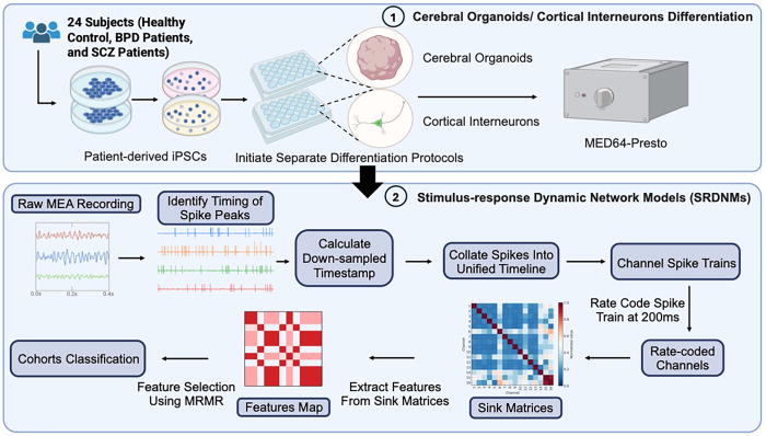
"Neuropsychiatric disorders such as schizophrenia (SCZ) and bipolar disorder (BPD) remain challenging to diagnose due to the absence of objective biomarkers, with current assessments relying largely on subjective clinical evaluations.
In this study, we present a computational analysis pipeline designed to identify disease-specific electrophysiological signatures from multi-electrode array (MEA) recordings of patient-derived cerebral organoids (COs) and two-dimensional cortical interneuron cultures (2DNs).
Using a Support Vector Machine classifier optimized for high-dimensional data, we achieved 95.8% classification accuracy in distinguishing SCZ from control samples in 2DNs under both baseline and post-electrical-stimulation (PES) conditions with the extracted electrophysiological signatures.
In COs, classification accuracy improved from 83.3% at baseline to 91.6% following PES, enabling robust separation of control, SCZ, and BPD cohorts.
Key discriminative features included channel-specific measures of network activity, with PES significantly enhancing classification performance, particularly for BPD.
These results underscore the potential of MEA-based functional phenotyping, coupled with machine learning, to uncover reliable, stimulation-sensitive electrophysiological biomarkers, offering a path toward more objective diagnosis and personalized treatment strategies for neuropsychiatric disorders."

Neural basis of schizophrenia and bipolar disorder found in brain organoids | Hub "Using machine learning, Johns Hopkins researchers identified healthy and unhealthy patterns based on electrical activity"


FIG. 1.The workflow of the proposed analysis pipeline was designed from EEG analysis to uncover distinct electrophysiological signatures associated with schizophrenia (SCZ) and bipolar disorder (BPD).


Wednesday, September 10, 2025

Researchers find how epilepsy genes disrupt different brain regions using stem cell models

Good news!

"Key takeaways
  • ... researchers used patient-derived stem cells to model how gene variants that cause developmental and epileptic encephalopathy type 13, a rare genetic childhood epilepsy, affect different regions of the brain.
  • ​​​​​​The team discovered that the same variants drive seizure-like hyperactivity in the cortex but disrupt memory-related neural rhythms in the hippocampus by depleting inhibitory neurons — offering insight into why seizure medications alone may not address the full scope of symptoms.
  • By reproducing abnormal brain activity observed in patients, the study establishes the first hippocampal assembloid model, creating a new platform for studying epilepsy, autism, Alzheimer’s disease and other brain disorders.
...

Using patient-derived induced pluripotent stem cells, the researchers generated advanced models known as 3D assembloids of two key brain areas:
the cortex, which is essential for movement and higher-order thinking, and
the hippocampus, which supports learning and memory. The results revealed strikingly different effects depending on the brain region.

In cortical models, the SCN8A variants made neurons hyperactive, mimicking seizure activity.
In hippocampal models, however, the variants disrupted the brain rhythms associated with learning and memory. This disruption stemmed from a selective loss of specific hippocampal inhibitory neurons — the brain’s traffic cops that regulate neural activity. 

These findings may help explain why patients with epilepsy often struggle with symptoms beyond seizures. ..."

From the highlights and abstract:
"Highlights
• Cortical assembloids with SCN8A mutations exhibit marked network hyperexcitability
• Hippocampal assembloids show theta-gamma coupling deficits, mirroring patient recordings
• Computational modeling predicts selective O-LM interneuron loss in the hippocampus
• scRNAseq and IHC reveal region-specific neuronal identity changes in DEE-13 assembloids

Summary
Neurodevelopmental disorders often impair multiple cognitive domains. For instance, a genetic epilepsy syndrome might cause seizures due to cortical hyperexcitability and present with memory impairments arising from hippocampal dysfunction.
This study examines how a single disorder differentially affects distinct brain regions using induced pluripotent stem cell (iPSC)-derived cortical- and hippocampal-ganglionic eminence assembloids to model developmental and epileptic encephalopathy 13, a condition arising from gain-of-function mutations in the SCN8A gene encoding the sodium channel Nav1.6.
While cortical assembloids showed network hyperexcitability akin to epileptogenic tissue, 
hippocampal assembloids did not, and instead displayed network dysregulation patterns similar to in vivo hippocampal recordings from epilepsy patients. Predictive computational modeling, immunohistochemistry, and single-nucleus RNA sequencing revealed changes in excitatory and inhibitory neuron organization that were specific to hippocampal assembloids.
These findings highlight the unique impacts of a single pathogenic variant across brain regions and establish hippocampal assembloids as a platform for studying neurodevelopmental disorders."

UCLA researchers find how epilepsy genes disrupt different brain regions using stem cell models | UCLA


Graphical abstract


Saturday, April 19, 2025

ALS Disturbs Sleep Prior to onset of ALS Symptoms and associated genetic mutations

This seems to be a plausible connection! Do not older people get less sleep and sometimes less quality sleep (e.g. reduced rapid eye movement or REM)?

Unfortunately, the study did not investigate whether sleep disturbances cause ALS. We are left with a kind of a chicken and egg problem.

Therefore, I also suspect, the pre onset sleep disturbances are not a good indicator for developing ALS.

This mix of sleep disturbances and genetic mutations make this study confusing. More research is probably needed.

"Sleep disturbances are commonly reported by people with neurodegenerative conditions that damage motor neurons. Some of these disorders, including amyotrophic lateral sclerosis (ALS), affect nonmotor functions of the brain, including sleep, which is regulated by the hypothalamus. ...

The researchers started by investigating differences in sleep quality between individuals who had early-stage ALS, a period before the onset of respiratory problems, and people without ALS or other motor deficits. They observed that people with ALS took longer to fall asleep and had increased bouts of wake and rapid eye movement (REM) sleep but reduced deep sleep non-REM (NREM) stages.

In a second cohort of individuals with an immediate relation to someone with ALS but who had not developed motor symptoms, the researchers recorded their sleep and collected DNA samples to determine which individuals carried a genetic mutation associated with ALS.

Compared to individuals with no ALS mutations, presymptomatic ALS carriers demonstrated altered sleep patterns that were dependent on the mutation that they carried: 
one mutation was associated with less overall time asleep and reduced deep sleep NREM, while a 
second mutation led to a longer time to fall asleep and more time in REM, but less time in total in NREM stages. Both mutations were associated with increased times in the wake period. ...

The team confirmed the sleep changes they saw in their human studies using three different animal models of ALS with mutations in either an RNA-binding protein, a DNA-binding protein, or superoxide dismutase, all shown to be affected in the disorder. Although the onset of sleeping changes varied by genetic mutation, the researchers saw an overall decrease in NREM and REM and increased wake periods in animals; these changes occurred prior to motor deficits in one model. Thus, although the effects to REM sleep differed between mice and humans, ALS caused similar changes to sleep-wake patterns in both species. ..."

From the editor's note and abstract:
"Editor’s summary
Patients with amyotrophic lateral sclerosis (ALS) often experience poor sleep, but whether these disturbances are caused by alterations in sleep regulating brain circuits or triggered by motor symptoms remains unclear.
Guillot et al. found that patients with ALS and presymptomatic carriers of ALS risk genes show increased wakefulness and reduced non–rapid eye movement sleep. Disruptions in sleep architecture were also found in three mouse models of ALS and partially normalized by intraventricular injection of melanin-concentrating hormone or oral administration of an orexin antagonist.
These results suggest that sleep disturbances occur before symptom onset and can be ameliorated by targeting neuropeptides involved in sleep/wake regulation. ...

Abstract
Sleep alterations have been described in several neurodegenerative diseases yet are currently poorly characterized in amyotrophic lateral sclerosis (ALS).
This study investigates sleep macroarchitecture and related hypothalamic signaling disruptions in ALS. Using polysomnography, we found that both patients with ALS as well as asymptomatic C9ORF72 and SOD1 mutation carriers exhibited increased wakefulness and reduced non–rapid eye movement sleep.
Increased wakefulness correlated with diminished cognitive performance in both clinical cohorts. Similar changes in sleep macroarchitecture were observed in three ALS mouse models (Sod1G86R, FusΔNLS/+, and TDP43Q331K).
A single oral administration of a dual-orexin receptor antagonist or intracerebroventricular delivery of melanin-concentrating hormone (MCH) through an osmotic pump over 15 days partially normalized sleep patterns in mouse models. MCH treatment did not extend the survival of Sod1G86R mice but did decrease the loss of lumbar motor neurons. These findings suggest MCH and orexin signaling as potential targets to treat sleep alterations that arise in early stages of the disease."

ALS Disturbs Sleep Prior to Symptom Onset | The Scientist "Humans and mice with a predisposition for ALS displayed altered sleep patterns before symptoms began, offering a potential novel diagnostic and treatment approach."

Friday, May 31, 2024

People with autism are 3X likelier to develop Parkinson’s-like symptoms

Bad news, if confirmed!

"People with autism are 3X likelier to develop Parkinson’s-like symptoms—5.98% compared to a rate of .11%-1.85% in the general population, according to a review of medical records for 247,539 people in the U.S.; the results have not yet been peer-reviewed."

Global Health NOW: The Kenyan Factory Producing Self-Reliance; A Forgotten, Yet Life-Threatening Infection; and ‘A National Crime Scene’

Autistic people three times more likely to develop Parkinson’s-like symptoms (original news release) Largest study of its kind also finds increased risk in older adults with a range of intellectual disabilities.

Saturday, January 13, 2024

Breakthrough Alzheimer's Discovery Reveals Five Distinct Variants

Good news! Getting the details right may make a huge difference!

"... The international research team that analyzed cerebrospinal fluid proteins in 606 people says this means that medications already tested may have falsely seemed to be ineffective or only slightly effective.

Their discovery could lead to more personalized therapies or preventive measures for these subtypes. It also presents hope for early diagnosis and intervention to delay the onset of symptoms of Alzheimer's disease (AD). ...
The researchers were able to identify five distinct biological subtypes of the disease, distinguished by variations such as hyperplasticity, immune activation, RNA dysregulation, choroid plexus dysfunction, and blood-brain barrier impairment. ..."

From the abstract:
"Alzheimer’s disease (AD) is heterogenous at the molecular level. Understanding this heterogeneity is critical for AD drug development. Here we define AD molecular subtypes using mass spectrometry proteomics in cerebrospinal fluid, based on 1,058 proteins, with different levels in individuals with AD (n = 419) compared to controls (n = 187). These AD subtypes had alterations in protein levels that were associated with distinct molecular processes: subtype 1 was characterized by proteins related to neuronal hyperplasticity; subtype 2 by innate immune activation; subtype 3 by RNA dysregulation; subtype 4 by choroid plexus dysfunction; and subtype 5 by blood–brain barrier impairment. Each subtype was related to specific AD genetic risk variants, for example, subtype 1 was enriched with TREM2 R47H. Subtypes also differed in clinical outcomes, survival times and anatomical patterns of brain atrophy. These results indicate molecular heterogeneity in AD and highlight the need for personalized medicine."

Breakthrough Alzheimer's Discovery Reveals Five Distinct Variants : ScienceAlert


Fig. 1: Biological description of AD subtypes.