Showing posts with label mammals. Show all posts

Friday, October 09, 2026

The development of mammal embryos, cell by cell

Amazing stuff!

I am not sure I like the expression "DNA typewriter". Obviously, a bad choice! When was the last time any of the involved researchers used a typewriter if ever?

"Decades ago, biologists traced the history of the thousand or so cells that create a nematode from single, fertilized cell.
Documenting such a developmental map for a mammalian embryo is much, much harder for various reasons. Two teams, however, have used genome editing tools in mouse embryonic stem cells or mouse zygotes to create the most comprehensive cell lineage maps so far for early mammalian development.

As the cells of a mouse embryo divided, the editors inserted DNA sequences that let researchers later discern their lineages. Revealed in parallel papers in Cell and Science , the studies chart cell relationships up to the point where the embryonic rodents’ organs begin to form. “  ...

The ultimate goal, a family tree tracing the ancestry of every cell in the mammalian body, could clarify how organs arise, probe the origins of birth defects, help grow better replacement cells for patients, and improve evolutionary studies."

"... To generate lineage maps for mice, both research teams used a DNA-modifying technique known as prime editing to insert distinctive marker sequences into the genomes of embryonic cell as development proceeded. From the modifications that descendant cells accumulated, the researchers could reconstruct how they were related. ..."

From the highlights and abstract (1):
"Highlights
• PEtracer resolves fate dynamics throughout early mouse embryogenesis
• >1.4 million deeply sequenced cells with ∼75% of cell divisions marked
• Quantitative view of lineage biases, restriction timing, and clonal outputs by tissue
• Highly reproducible lineage architecture across independent replicate embryos

Summary
A comprehensive cell fate map of mammalian embryogenesis has remained out of reach given the scale, cellular diversity, and non-deterministic nature of development in utero.
Here, we use PEtracer to continuously install heritable genetic marks as development progresses, reconstructing lineage trees that resolve ∼75% of cell divisions across >1.4 million cells from 16 replicate embryos.
We pair these trees with deep transcriptional profiling to resolve cell fate biases, restriction timing, progenitor pool sizes, and lineage relationships throughout embryogenesis.
Using this quantitative reference, we uncover strikingly reproducible lineage architecture between replicate embryos and chart the lineage dynamics driving fate specification across diverse tissues.
Specific biological insights include the progressive restriction of neural crest fate, the relative contributions of distinct mesodermal origins to endothelium, and the dynamics of axial elongation. This work provides a foundation for a quantitative and predictive understanding of mammalian development."

From the abstract (2):
"Mammalian biology unfolds over time, within tissues and organs opaque to our eyes and instruments.
We applied DNA Typewriter, a sequential molecular recorder, to record the cell lineage of a mouse over nearly two weeks of development.
From one embryo, we reconstruct a time-calibrated, parsimony-supported, zygote-rooted phylogeny of 1.28 million transcriptionally profiled cells.
A burst of editing unequivocally marks the daughters of the first cleavage, which serve as inline replicates.
We quantify clonal dominance arising during gastrulation.
Tree siblings share cell type far above chance; heterotypic siblings mark terminal differentiations. Temporal sweeps of clade co-occurrence recover a dated hierarchy of cell-type couplings; imputed labels for internal nodes recapitulate known state paths. A lineage-anchored ontogeny of mammalian development, long out of reach, is coming into view."

ScienceAdviser


Graphical abstract (1)


Figure 1 Embryonic-scale cell fate mapping of mouse development with PEtracer (1)


Figure 1. Pronuclear zygotic injection (PNI) of DNA Typewriter components identifies an E13.5 embryo with robust levels of sequential editing. (2)


Monday, September 28, 2026

Elemental sulfur found in mammalian cells for first time

Amazing stuff!

"Mammalian cells actively produce elemental sulfur, using it as an antioxidant to limit cell damage. While bacteria, plants and fungi are known to produce the material, this is the first time it has been detected in mammals. ...

Now, an international team has detected S8 in cells from both mice and humans. The team, ... used a polyaromatic capsule to trap and stabilise S8 through hydrophobic interactions. This allowed them to use Raman microscopy and mass spectrometry to locate and quantify the abundance of elemental sulfur. ..."

"... Antioxidants are not obtained exclusively through the diet, as the body can also synthesize them with the help of various enzymes. An international research team ... has now demonstrated a new mechanism that contributes to the body’s own production of antioxidants. The researchers showed that mammalian cells produce and store elemental sulfur in a controlled manner in the form of cyclooctasulfur (S₈), a molecule consisting of eight sulfur atoms.

At the heart of this protective system is the enzyme eNOS, which is found in many cells. It produces nitric oxide (NO), a molecule that relaxes blood vessels and thereby regulates blood pressure. The researchers have now shown for the first time that eNOS has another function: the enzyme produces cyclooctasulfur (S₈), which serves as a storage form of elemental sulfur in cells. ...

The researchers found particularly high concentrations of these “sulfur rings” in the oxygen-dependent mitochondria, the powerhouses of the cell, as well as in lipid droplets, which serve as the cell’s fat stores. Through chemical reactions, the body can use its supply of S₈ molecules to produce specialized sulfur compounds known as hydropersulfides, which act as antioxidants. ..."

From the editor's summary and abstract:
"Editor’s summary
Elemental sulfur is an inert, water-insoluble form of sulfur that is converted into biologically assimilated forms used in energy metabolism, especially in bacteria in plants. Barayeu et al. report that cyclo-octasulfur is present in millimolar amounts in mitochondrial membranes and lipid droplets in human and mouse cells ... 
Octasulfur was generated by endothelial nitric oxide synthase. This process may provide a storage source for reactive hydropolysulfides that allow antioxidant defense mechanisms in mammalian cells. Supporting such a protective role, depletion of octasulfur in cultured mouse cells increased lipid peroxidation and cell death. ...

Structured Abstract
INTRODUCTION
Elemental sulfur is a central intermediate in the global sulfur cycle and is widely produced by prokaryotes, with additional evidence for its use in plants and fungi. Whether mammals synthesize, store, or use elemental sulfur, however, has remained unknown. Because mammalian cells enzymatically generate diverse reactive sulfur species, including hydropersulfides and polysulfides, we asked whether they also produce cyclo-octasulfur (S8), the most stable allotrope of elemental sulfur, for biological function.

RATIONALE
We reasoned that if mammals use elemental sulfur as a biologically meaningful metabolite, S8 should be maintained not as an incidental chemical product but as a regulated intracellular reservoir coupled to reactive sulfur metabolism.
To test this possibility, we established complementary approaches:
mass spectrometry using a polyaromatic capsule that captures and stabilizes S8, enabling its quantitative analysis, and 
Raman microscopy for the direct visualization of S8 in mammalian cells and tissues.

RESULTS
In line with established knowledge, we detected S8 in bacteria and yeast and further found it in mammalian mitochondria and lipid droplets from mouse and human cells and tissues. S8 was especially enriched in lipid droplets, corresponding by extrapolation to tens of grams or more of S8 across the human body, with particularly high levels in adipocyte-rich breast cancer tissues.
We identified lipid droplet–associated endothelial nitric oxide synthase (eNOS) as a major source of S8. Recombinant eNOS converted glutathione polysulfides [e.g., glutathione trisulfide (GSSSG)] into longer sulfur chains and ultimately S8 through an NADPH-dependent sulfur-catenation reaction, where NADPH is the reduced form of nicotinamide adenine dinucleotide phosphate; in the presence of glutathione polysulfides and the canonical eNOS substrate L-arginine, this reaction also generated the nitric oxide (NO) carrier S-nitrosoglutathione (GSNO). Functionally, depletion of endogenous S8 increased lipid peroxidation and ferroptotic cell death,
whereas S8 delivery protected adipocytes and endothelial cells from ferroptosis. Mechanistically, S8 reacted with abundant cellular thiols, such as glutathione (GSH), to generate antioxidant hydropersulfides and polysulfides that suppress lipid peroxidation.
Local delivery of solubilized S8 into joints of mice with experimental osteoarthritis also reduced lipid peroxidation in joint tissue, suggesting a potentially protective effect.

CONCLUSION
Our findings establish that mammalian cells not only synthesize elemental sulfur as S8 but also sequester it within lipid-rich compartments at concentrations that surpass those of canonical lipophilic antioxidants such as vitamin E.
This hydrophobic sulfur reservoir may provide a dynamic replenishment of redox-active persulfides, thereby linking sulfur-chain chemistry to eNOS-dependent signaling, the restraint of lipid peroxidation, and resistance to ferroptotic cell death. These results position elemental sulfur biosynthesis as an evolutionarily conserved yet metabolically adaptable antioxidant strategy, broadening the conceptual landscape of mammalian redox biology."

Elemental sulfur found in mammalian cells for first time | Research | Chemistry World

Fighting cell death with sulfur (original news release)


A polyaromatic capsule was used to trap sulfur molecules produced in mammalian cells


Mammalian biosynthesis of elemental sulfur.


Friday, September 18, 2026

Weeping whales: Stillborn humpback whale grieving documented

Amazing stuff!

"Humpback whale mothers may experience grief when their calves die, a rare research observation led by Griffith University and Sea World Foundation has found.

In 2025, a female humpback whale—along with an escort whale—was observed off the southern Gold Coast after giving birth to a presumed deceased calf. She remained with it for several hours and possibly even days.

Dr. Olaf Meynecke, from Griffith University's Whales and Climate Research Program, said postmortem attentive behavior in cetaceans had been documented predominantly among toothed whales and delphinids (known as odontocetes), but remained largely absent from research published on baleen whales, like humpback whales. ...

“She remained close to the calf, maintaining eye contact and positioning herself next to her calf over several hours and maybe even days. ..."

Weeping whales: Stillborn humpback whale grieving documented


These images show the stillborn calf and mother, and amniotic membrane just after the birth


Wednesday, September 16, 2026

Wild dogs make the longest recorded journey of any terrestrial mammal in Africa

Amazing stuff!

"... Researchers from the Zambian Carnivore Programme and Montana State University wanted to understand how this endangered predator navigates human-dominated landscapes to better help with conservation efforts. So they tracked groups of wild dogs in Zambia after they separated from their packs.

In doing so, they recorded a record-breaking journey. Three of the dogs traveled a staggering 4,126 kilometers (2,564 miles) across the country, the longest journey ever recorded for an African land mammal. ..."

Wild dogs make the longest recorded journey of any terrestrial mammal in Africa


African wild dog (Lycaon pictus) in the Okavango Delta, Botswana.


Monday, September 14, 2026

Bats carry dangerous viruses without getting sick

Amazing stuff!

"Scientists discovered that more than 500 species of vesper bats possess two separate sets of antibody genes, an immune arrangement never before found in mammals. The surprising system could help explain how bats tolerate so many viruses without becoming seriously ill."

"... that the world’s largest family of bats has two distinct copies of the genes that produce antibodies, the specialized proteins that help the immune system recognize and fight infections. Every other known mammal has only one such set. ..."

From the abstract:
"Bats are major reservoirs of viruses that can be transmitted to humans in zoonotic outbreaks.
Antibody-mediated immunity plays an important role in shaping viral evolution and immune evasion but remains understudied in bats.
All known mammals have a single immunoglobulin heavy chain (IgH) gene locus and up to two light chain loci.
We have identified dual IgH loci on separate chromosomes in 26 bat species, highlighting extreme variation of immunogenetic architecture in order Chiroptera. In a model species, Eptesicus fuscus, we leveraged single-cell transcriptomes to confirm functional rearrangement and expression of both loci, but with different mechanisms for generating antibody diversity and function. These results provide a foundation for analysis of humoral immunity and pathogen response in bats."

Global Health NOW: Where Measles Finds Footholds; and How Climate Change Accelerates AMR


Fig. 1. Genomic annotations and analysis for both IGH loci.


Thursday, September 10, 2026

Males are more difficult to birth than females for many mammals. Really!

Amazing stuff! This about the obstetrical dilemma. Was it not known for thousands of years that male babies are on average larger than female babies and thus impose more risks during childbirth?

Apparently, the only new discovery here is that these risks are found across mammal species.

I am not sure why the AAAS (American Association for the Advancement of Science) picked this article to promote it!

Again the AAAS gets caught using the ideological term "people" instead of women! DEI demagoguery is still alive!

The costly son hypothesis is a very unfortunate term. I hope it will be quickly end up in the dustbin of history and gain no traction!

"Every day, about 700 [women] people die giving birth around the world. A leading cause is obstructed labor, often due to the baby’s head exceeding the size of the birth canal. ...

Reviewing birth data for 140 species, the authors argue that sons are more difficult to birth than daughters for many mammals, including humans and our ancient ancestors.
The authors found this pattern in five domesticated animals: cattle, water buffalo, sheep, goats, and a breed of American racehorse.
That’s also the case in captive antelope and Asian elephants.
The pattern holds in cattle and sheep, even when controlling for birth weight, suggesting in addition to sheer size, factors such as sex differences in fetal shape and behavior could contribute to males causing more trouble during birth. ...

This added risk of this phenomenon, which the researchers dubbed the “costly son” hypothesis ..."

From the abstract:
"Human childbirth has often been considered uniquely difficult among mammals, yet recent evidence suggests that obstructed labor and fetopelvic disproportion are widespread across placental mammals.
Here, we propose the “costly son” hypothesis: because male offspring are typically larger at birth, they impose greater obstetric risk on mothers and themselves by increasing the likelihood of fetopelvic disproportion and dystocia. We compiled sex-specific birth weight data for 140 placental mammal species across 11 orders and reviewed published evidence on sex differences in dystocia risk.
Male neonates were more commonly larger at birth than females, and male-biased birth size differences were strongly associated with male-biased adult sexual size dimorphism. Species with male-biased adult size dimorphism had sixfold greater odds of exhibiting male-biased birth weight differences than species with female-biased dimorphism.
Male-biased birth weight differences existed irrespective of developmental mode (precocial or altricial), indicating that sex differences in fetal growth arise early in gestation.
Evidence from humans, livestock, and at least one non-human primate suggests that male fetal sex is associated with elevated dystocia risk, that is, precocial taxa where birth size approaches the mechanical limits of the maternal birth canal.
We further hypothesize and discuss how parity and sex ratio adjustment may modulate these risks and associated trade-offs in a Trivers–Willard context.
Our findings identify fetal sex as an underappreciated dimension of mammalian obstetrical dilemmas and suggest that the evolutionary benefits of producing larger sons are balanced by increased risks of birth complications and mortality."

ScienceAdviser

Thursday, September 03, 2026

Scientists identify early signal of dangerous pregnancy complications

Good news! This could be a breakthrough!

"In a study published today [8/31/2026] ... scientists who analyzed blood samples from thousands of pregnant women found that low levels of a protein called ISM2 during the first trimester were associated with higher risk of both preeclampsia and fetal growth restriction later on.
Follow-up lab studies of placental cells showed ISM2 likely plays a causal role in the disorders, meaning the findings could help in the development of treatments, too. ...

The researchers measured blood levels of nearly 3000 proteins and found one in particular associated with both preeclampsia and fetal growth restriction: ISM2, a little-studied protein produced by the placenta. Specifically, lower blood levels of the protein at the 12-week blood test were linked to higher risks of preeclampsia, fetal growth restriction, or both. ...

What’s more, ISM2 levels were a better predictor of each condition than currently used proteins. ...

In lab experiments, the team found that blocking the production of ISM2 in stem cells that usually develop into normal, uterus-invading EVTs stopped that process from happening.
By contrast, artificially adding ISM2 into a cell line that usually lacks it induced recipient cells to migrate around lab dishes more like EVTs would. ..."

"... Both of these conditions are known to be linked to problems with the placenta not developing properly early in pregnancy, in particular a failure of specialised placental cells known as the extravillous trophoblast (EVT) to invade the mother’s womb and establish a healthy blood supply. ...

Of all mammals, humans have the deepest placental invasion, which reflects the massive demands of oxygen and nutrients required to manufacture the most complex machine ever known, the human brain. ..."

From the abstract:
"Preeclampsia and fetal growth restriction (FGR) are major causes of global morbidity and mortality. 
Both conditions are associated with impaired invasion of the uterus by extravillous trophoblast (EVT).
We performed proteomics in maternal serum obtained at ~12 weeks of gestational age in a prospective pregnancy cohort (Pregnancy Outcome Prediction Study). Here we show that low maternal serum isthmin-2 (ISM2) was the strongest protein signal (out of 2,904) in the first trimester of pregnancy for preeclampsia or FGR.
We validated the association in two independent cohorts (Pregnancy Outcome Prediction Study 2 and Improving Maternal Pregnancy And Child ouTcomes study).
ISM2 protein and mRNA are almost exclusively produced in the placenta, and, within the placenta, ISM2 mRNA is highly enriched in EVT. 
Knocking down ISM2 in cultured human trophoblast stem cells profoundly inhibited EVT invasion.
Conversely, expressing ISM2 in a cell line lacking endogenous ISM2 (HEK293 cells) promoted migration.
We conclude that ISM2 may be causally involved in the early pathophysiology of failed trophoblast invasion and that the protein and its associated pathways are potential targets for the prediction and prevention of preeclampsia and FGR."

Scientists identify early signal of dangerous pregnancy complications | Science | AAAS "Low levels of a placenta made protein predict preeclampsia and fetal growth restriction and could offer new target for prevention"

Pre-eclampsia discovery could lead to new test and treatments for at-risk pregnancies (original news release) "Cambridge scientists have identified a key protein that could act as a predictor for pre-eclampsia and fetal growth restriction, two of the major causes of stillbirth and of sickness among mothers and newborns."


Fig. 2: The association between ISM2 and PE and FGR.


Fig. 3: ISM2 regulates EVT differentiation.


Saturday, August 29, 2026

Key brain circuit regulating the entry into hibernation found at last

Amazing stuff!

Notice here the emphasis is on entry into hibernation not hibernation in general.

Notice Google Search was not able to find a news release regarding this discovery neither did the Whitehead Institute for Biomedical Research publish one although many of the authors of this study are affiliated with the WI. That is disappointing.

"... Now, in a preprint posted on bioRxiv, researchers studying hibernating Syrian hamsters have pinpointed for the first time a brain circuit that regulates an animal’s entry into hibernation. The results reveal a “key, really, in the network” and provide “more solid [evidence] than anything we had before” of an ancient hibernation circuit within the mammalian brain ..."

From the abstract:
"Evolution of seasonal hibernation has enabled mammals to survive harsh conditions by entering a state of prolonged hypometabolism and hypothermia with body temperatures as low as 0-4°C1–6.
Despite decades of physiological studies, the genetic tools to study hibernation have remained limited and the mechanisms that induce hibernation entry are still unknown.
Focusing on the brain, we map state-dependent neuronal activity across the hibernation cycle in Syrian hamsters and identify the hypothalamic anterior preoptic area (aPOA) as a key regulator of hibernation entry.
Single-nucleus RNA and chromatin profiling provided a map of neuronal populations present in the hamster POA and enabled the discovery and design of an enhancer AAV that selectively targets hibernation-associated aPOA subpopulations.
Using this genetic approach, we show that Samd3-positive aPOA glutamatergic neurons are necessary for entry into hibernation and that their activation is sufficient to induce a prolonged hypothermic state with associated nesting behavior.
Together, we identify the first neuronal population that controls entry into hibernation, opening new avenues for investigating and manipulating the metabolic and physiological mechanisms underlying this extreme state of “suspended animation” and its potential applications in aging and disease."

Key brain circuit regulating hibernation found at last | Science | AAAS


Fig. 2: Identification of anterior preoptic area activity as necessary for deep torpor entry.


Saturday, August 01, 2026

How did mammals from North America move into South America millions of years ago

Amazing stuff!

"... researchers have found fossil evidence that animals from different latitudes of North America and South America mixed long before the Panamanian land bridge permanently linked the two continents about 3 million years ago.
Since around 10 million years ago, mammals — primarily small ones like sloths and coatis — found a way across the watery barrier. Only larger mammals had to wait for the permanent land corridor. ...

Biologists thought that only after the isthmus formed did animals surge north and south, what biologists refer to as the Great American Biotic Interchange or GABI. That resulted in the extinction of a lot of South American species as North American species took over.

Yet this picture of mammal movements across continents is too simple, ... paleontologists argue. After assembling and analyzing a new database of fossil mammals discovered in Mexico, they conclude that northern North American animals had already migrated into southern North America by about 10 million years ago, where they waited around in a kind of holding pen until opportunities arose for some of them to continue crossing to the south starting about 7 million years ago. ...

During that pre-GABI time, smaller mammals — heralded by racoon-like coatis and ringtails and small rodents — moved to South America via some kind of intermittent corridor, by island hopping through the Caribbean or by rafting on vegetation. Only one major South American group, the sloths, moved to North America. ..."

From the abstract of the Perspective:
"Whenever plants and animals newly come together, there will be some winners and some losers. This was the case for the Great American Biotic Interchange (GABI), which occurred ~2.8 million years ago (Ma), when the emergence of the Isthmus of Panama connected the continents of South and North America. Although species moved both northward and southward during the GABI, a massive taxonomic asymmetry arose between the two continents, with a much greater proportion of species in South America having a North American origin than the opposite. ... Tseng et al. report that a large fossil dataset from Mexico helps to uncover the events that primed mammals for movement across the Isthmus."

From the editor's summary and abstract:
"Editor’s summary
For millions of years after the breakup of the supercontinent Gondwana, South America was disconnected from other landforms, a time period that has been referred to as its “splendid isolation.”
This isolation allowed for the evolution of mammals with distinctive features, including xenarthrans, sprassodont carnivores, and notoungulates.
The formation of the Panamanian land bridge facilitated connection of North and South American faunas, with a bias from north to south. Tseng et al. looked at fossils from low latitudes in México and found that northern mammals accumulated in the region before the connection (see the Perspective by McGuire and Williams). This allowed for more rapid dispersal south and further supports that the land bridge first formed during the Pliocene rather than the Miocene. ...

Abstract
The movement of taxa between North and South America across the Panamanian land bridge, known as the Great American Biotic Interchange (GABI), established modern-day mammal assemblages in the Americas, heralding the decisive end of what has been called the “splendid isolation” of the South American fauna.
Lacking substantive low-latitude records, previous work on this pattern relied on South American and temperate North American fossils to conclude that pulsed interchanges began about 2.8 million years ago (mya).
Based on new low-latitude fossil data from México, we found evidence for a prolonged, triphasic interchange.
Phase 1 consisted of the initial accumulation of northern higher-latitude mammals in a Mexican “holding pen” beginning 10 mya. 
Dispersal began in phase 2, with a strong north-south gradient and increasing biogeographic connections (7 to 3 mya).
The final phase consisted of the already well-described taxonomic pulses (from ~2.8 mya)."

ScienceAdviser

When mammals crossed between continents (no public access)

North and South America weren’t always connected, but their mammals still found a way to intermingle (original news release) "Before the Panamanian land bridge formed about 3 million years ago, mammals on the two continents were isolated — or so biologists thought. New fossils show that they found ways to mix long before that."

A 10-million-year biogeographic holding pen primed the Great American Biotic Interchange (no public access) "Before the Panamanian land bridge formed about 3 million years ago, mammals on the two continents were isolated — or so biologists thought. New fossils show that they found ways to mix long before that."


This illustration depicts (left) large North American mammals — counterclockwise from top, mammoths, llamas, tapirs, peccaries and saber-toothed cats — waiting in a kind of holding pen in Mexico for the bridge to form.
In South America, the mammalian interchange involved, counterclockwise from top, ground sloths, marsupials, capybaras, armadillos, glyptodonts and, incidentally, terror birds. While North American mammals took over in South America, only about 10% of North American mammals today are derived from mammals that originated in South America.


Wednesday, April 22, 2026

Researchers have imaged every single cell in the body of a newborn mouse

Amazing stuff! 

"Researchers have imaged every single cell in the body of a newborn mouse, using a custom fluorescence microscope, high-speed cameras and an atlas of the mouse body onto which they mapped data. 

“Since Robert Hooke first described cells in 1665, more than 360 years have passed,” ... “Yet no one had directly visualized all cells in mammalian organs or in the whole body of a newborn mammal at single-cell resolution.” They also mapped responses to drug toxicity at a single-cell scale."

Nature Briefing: Translational Research

Establishing whole-body cellomics (partially behind paywall)


Friday, March 27, 2026

Unprecedented footage shows sperm whales joining forces to help a newborn calf

Amazing stuff!

"The researchers quickly realized they had in fact observed a group of whales helping a calf take its first breath. Even more fortuitous, they had captured it on video from a drone.
It’s rare for researchers to see a sperm whale birth, with only a handful having been recorded previously.
And this time, overhead footage allowed the scientists to meticulously analyze the individual whales’ behavior before, during, and after the delivery. Although researchers have long hypothesized that sperm whales cooperate in certain social situations, the new study—reported today in Science—shows whales from different family branches can work together to support a newborn calf. ..."

Unprecedented footage shows sperm whales joining forces to help a newborn calf | Science | AAAS

Wednesday, March 25, 2026

Study suggests healing skin without scarring may be possible

Good news!

"... The new study published March 20 in Cell reveals a way to fully regenerate skin by unblocking an embryonic healing mechanism that shuts off after birth. Demonstrated on mice, the strategy may help guide the development of similar therapies for human patients. ...

In mice wounded three days before birth, the skin regenerated diverse cell types and closely resembled unwounded skin.
But when wounded at five days after birth, the site was covered by epithelial cells and became packed with collagen scar tissue and abnormally dense nerve fibers and immune cells. Many other skin cell types failed to regrow. ...

They found that postnatal wound sites became densely packed with nerves. This “hyperinnervation” occurs because fibroblasts in postnatal wounds upregulate the gene Cxcl12, which recruits excessive nerves to the area and impairs the regrowth of other skin-cell types.

When researchers depleted Cxcl12 in wounds in postnatal mice, “hyperinnervation” was curtailed, and the skin regrew diverse cell types. Blocking local nerve signaling with botulinum toxin A (Botox) produced similar effects. ..."

From the highlights and abstract:
"Highlights
• Embryonic skin regenerates diverse cell types after injury, whereas postnatal skin does not
• Postnatal wounds have wound-specific fibroblasts that drive hyperinnervation via CXCL12
• Excessive innervation at the wound site blocks multilineage regeneration
• Reducing hyperinnervation restores multilineage regeneration after postnatal injury

Summary
Some mammalian tissues can replace lost cells within one lineage, but organ-level regeneration—restoring diverse cell types across lineages—remains rare. Here, we show that late embryonic full-thickness skin injuries heal by regenerating epithelial, mesenchymal, neuronal, and vascular tissues with proper connectivity. However, this ability is lost soon after birth, resulting in failure to restore most cell types and hyperinnervation within the wound bed. Single-cell sequencing identified a postnatal wound-specific fibroblast (PWF) population absent after embryonic wounding. Through an in vivo screen, we discovered that three PWF-enriched genes—Timp1, Cxcl12, and Ccl7—inhibit organ-level regeneration and cause hyperinnervation when overexpressed in embryonic wounds. Reducing hyperinnervation in postnatal wounds through the depletion of Cxcl12 in fibroblasts or nerve ablation enables regeneration of diverse lineages after injury. Our study identifies mechanisms that transition an organ from regenerative to non-regenerative, discovers fibroblast-driven hyperinnervation as a key barrier, and demonstrates that removing this barrier unlocks organ-level regeneration.
"

Study suggests healing skin without scarring may be possible — Harvard Gazette "Researchers unblock embryonic regrowth mechanism that shuts down after birth in mice"


Graphical abstract


Figure 1 Multilineage regeneration differs between embryonic and postnatal wounding

Saturday, March 14, 2026

The cellular switch that explains why humans aren’t nocturnal

Amazing stuff! How mammals became diurnal after nocturnality!

"Early mammals were nocturnal, sleeping during the day while large predators were active. However, after the extinction of dinosaurs, several different lineages of mammals independently transitioned to become active during the day. Exactly how this dramatic change occurred has proved elusive. A new study, published in the journal Science, has revealed a cellular switch which holds the answer. ...

he study looked at how cells from a range of nocturnal (active at night) and diurnal (active in the day) mammals, like humans, respond to environmental signals.

Changes in temperature or osmolarity, as happens to the body throughout the day, caused the cells to respond in opposite ways, including in fundamental cellular functions. This divergence flips the timing of cellular activity, essentially acting as a day/night ‘switch’ at a molecular level.

The researchers pinpointed these differing responses to the mechanistic Target of Rapamycin (mTOR) and With-no-lysine (WNK) kinase pathways, central signalling networks in cells responsible for regulating several key functions, including protein synthesis. This suggested that modification of their activity could enable nocturnal mammals to switch to more diurnal activity.

To explore this hypothesis, the researchers administered diet-based treatments to mice to target the mTOR pathway, as mTOR activity is highly sensitive to nutrient levels. 
Once mTOR function was reduced, the mice began behaving more like diurnal animals, shifting their active hours into the daytime. This underlined that mTOR signalling goes beyond influencing metabolism; it also helps dictate when an animal is active. ..."

From the editor's summary and abstract:
"Editor’s summary
When ferocious dinosaurs roamed the earth, it was advantageous for mammals to be nocturnal. After these predators became extinct, many mammals (including the ancestors of humans) switched their daily rhythms to be active during the day. Beale et al. explored changes that might allow animals that retain the same fundamental clock components to make this switch. One cue that influences the timing of cellular clocks is temperature. Cells of nocturnal animals were more sensitive to temperature changes, and their clocks ran faster at higher temperatures. This appeared to reflect differential sensitivity of signaling pathways that regulate rates of protein translation through changes in protein phosphorylation. Inhibition of one pathway containing the protein kinase mTOR (mechanistic target of rapamycin) shifted nocturnal mouse cells toward more diurnal activity.  ...

Structured Abstract
INTRODUCTION
The ancestors of modern mammals were strictly nocturnal, avoiding the daytime while dinosaurs dominated. Only after the extinction of nonavian dinosaurs did mammals radiate into daytime niches, meaning that daily physiological rhythms became reversed with respect to the day and night but without any change in the brain’s master circadian clock.
Diurnality evolved multiple times, independently, but can also arise spontaneously in certain nocturnal species under conditions of very low energy balance. No specific neuroanatomical circuit has been demonstrated to function as a nocturnal-diurnal switch in any mammalian lineage, so we tested the hypothesis that this daily biological signal inverter instead has a cell-autonomous basis.

RATIONALE
Circadian timing is intrinsic to most mammalian cells. Daily hormonal cues, such as glucocorticoid and insulin signaling, potently synchronize cellular clocks with each other and the external day:night cycle, with very similar effects on the cellular clocks of diurnal and nocturnal mammals. However, the rates and equilibria of several fundamental biochemical processes were recently revealed to differ markedly between human and mouse cells. We therefore asked whether daily systemic rhythms (temperature, osmolality) that directly affect cellular biochemistry might elicit different effects on the function of diurnal versus nocturnal mammalian cells. To test this, we compared responses of cells and tissues under acute, long-term, and cyclical stimuli at the levels of circadian timing (bioluminescence reporter assays), proteins (proteome), protein modifications (phosphoproteome), and protein synthesis. We used comparative genomics to identify genes evolving with diurnal niche preference, and we perturbed candidate pathways in cells, tissues, and live mice to test causality.

RESULTS
Using daily thermal or osmotic cycles as a tool, we found opposite entrainment of diurnal versus nocturnal cells, reflecting their species’ temporal niche. Mouse and human cells differed not only in the magnitude but also the direction of their response to temperature. Moreover, temperature change evoked opposite shifts in global protein synthesis and phosphorylation between human and mouse cells and implicated differential sensitivity of the mechanistic target of rapamycin (mTOR) and with-no-lysine (WNK) kinase signaling pathways as plausible mediators.
Comparative genomics validated that genes in these pathways show accelerated evolution in diurnal mammals, rendering diurnal cells more robust against perturbations of solvent thermodynamics and consistent with an energy-saving adaptation. Last, manipulation of mTOR signaling in nocturnal mouse cells and tissues and in vivo was sufficient to shift circadian timing toward being more diurnal.

CONCLUSION
We identified a cell-intrinsic, thermodynamic mechanism underlying the mammalian switch between nocturnal and diurnal activity. By linking cellular responses to thermodynamic perturbation with circadian entrainment through genetic alterations in the mTOR and WNK pathways, our findings explain how diurnality could repeatedly evolve in mammals. More broadly, they highlight that even fundamental cellular properties, such as the response to temperature, may differ systematically between species, with profound consequences for circadian biology and temporal niche."

The cellular switch that explains why humans aren’t nocturnal | University of Cambridge "Differences in cellular pathway activity flip the switch from nocturnality to diurnality and explain a major evolutionary change humans have undergone."


Convergent evolution of cellular clock robustness to perturbation as a route to mammalian diurnality.


Sunday, February 15, 2026

Deer uses photoluminescence to mark trees like signposts

Amazing stuff!

"... So, to get noticed, deer mark areas with rubs and scrapes, known as signposts. Now, a new study in Ecology and Evolution reports that these signposts hold a hidden glow, and other deer can see it.

In the study the researchers describe this hidden glow as photoluminescence, the process by which an organic object absorbs light of a short wavelength and re-emits it at a longer wavelength. ...

The team observed the signposts glowing brighter during the breeding season. The scientists hypothesize more intensive rubbing generates this brighter glow. ..."

From the abstract:
"Ultraviolet (UV) induced photoluminescence is widespread in Mammalia; however, its function(s) remain unclear. Most of the research to date has focused on the surface expression of photoluminescence (e.g., pelage), described qualitatively.
Here, we report a quantitative assessment of photoluminescence of white-tailed deer (Odocoileus virginianus, herein deer) used for marking signposts. We analyzed 146 signposts, including 109 antler rubs on trees and 37 scent-marking scrapes. We compared the spectra of signposts to the spectra of surrounding environmental features elicited by exposure to excitation lights peaking at 365 and 395 nm. Signposts showed significant contrast when compared to environmental backgrounds (p < 0.001), and the resulting photoluminescence would be visible to deer based on previously described deer visual capabilities.
This research is the first quantitative description of the functional use of environmental photoluminescence by a mammal and gives new perspective about how white-tailed deer perceive their environment and communicate."

Deer communicate with hidden glow on signposts "The study is the first to unequivocally show how deer use a kind of photoluminescent marking system to communicate with one another."


The study is the first to unequivocally show how deer use a kind of photoluminescent marking system on trees to communicate with one another


Monday, January 05, 2026

Genomic study reveals how people moved pigs across the Asia-Pacific region for 50,000 years

Amazing stuff! The advantages of being an omnivore! 😊

"A new study ... reveals how millennia of human migration across Pacific islands led to the introduction of invasive pig species all over the Asia-Pacific region. ...

One notable exception is pigs. Pig populations occur on both sides of the Wallace Line and extend across Southeast Asia to New Caledonia, Vanuatu and remote Polynesia. Pigs are highly effective ecological invaders, and they are also culturally important across the region, raising a key question: what role did people play in their spread?

The new paper looked at the genome of over 700 pigs, including from living and archaeological specimens. This allowed the reconstruction of their movement across Southeast Asia and identify when they arrived on certain islands and how they might have interbred with various native pig species.

The researchers found that people of different cultures have moved pig species in the region for millennia. The earliest evidence points to people living in Sulawesi perhaps as early as 50,000 years ago, known to be the earliest cave painters, who both depicted and transported warty pig species as far away as Timor, possibly to establish future hunting stock.

The introduction of pigs in Island Southeast Asia dramatically accelerated, around 4,000 years ago, when early agricultural communities transported domestic pigs into the region.

Their journey began from Taiwan, extending across the Philippines, northern Indonesia (Maluku), into Papua New Guinea, and on to the outlying islands as far as Vanuatu, and remote Polynesia. The authors also found evidence for the introduction of pigs from Europe during the colonial period.

Many of these domestic pigs escaped, and became feral, in some cases, like on the Komodo islands, hybridizing with the warty pigs brought by people from Sulawesi thousands of years earlier. These hybrid pigs are now a major source of food for the endangered Komodo dragons.

The findings of this study highlight the dramatic and enduring impact of human activity on local ecosystems in the Pacific, raising conservation conundrums. Pigs in the region today have dramatically different statuses and impacts across islands: some are considered spiritual beings, others pests, while some are now so ingrained in local ecosystems that they could almost be considered native. ..."

From the editor's summary and abstract:
"Editor’s summary
Multiple species of pigs are endemic to Maritime Southeast Asia. Some of them exhibit a distinctive tooth shape and mitochondrial DNA haplogroups, but the history of pigs in this region is not clear. Stanton et al. collected genetic data from 117 modern, historic, and ancient individuals and examined morphometric data from over 700 modern and archeological samples. After comparing with 585 published mitochondrial genomes, the authors found that domestic pigs, which were likely introduced by Austronesian-speaking groups, contributed to most pigs found today from Hawaii to the Philippines. However, early specimens from these groups show no admixture with endemic species, mirroring the lack of mixture between these early human groups with local groups during their initial migrations. ...

Structured Abstract
INTRODUCTION
Humans have long moved animal species beyond their native ranges, drastically altering ecosystems, especially on islands. The translocation of vertebrates eastward across the Wallace Line into Wallacea, a biogeographic region between the Asian and Australasian biotas, has had major environmental impacts. For example, although the natural range of the genus Sus (pigs) is primarily located in Eurasia and Western Indonesia, free-living pigs are now widespread in Wallacea.

RATIONALE
The human-mediated translocation of pigs east of the Wallace Line most likely began with S. celebensis, a species native to Sulawesi, in pre-Neolithic times >4000 years ago.
S. scrofa (the ancestor of modern domestic pigs) populations were also likely introduced east of the Wallace Line between 4000 and 3000 years ago during the expansion of Austronesian speakers from mainland East Asia and Taiwan, through the Philippines and northern Wallacea, into Melanesia and Polynesia. The geographic origin and ancestry of these populations from Wallacea and Oceania, however, remain unknown.
To establish the geographic origins of the pig populations in Wallacea, Melanesia, Micronesia, and Polynesia and the potential dispersal routes along which they traveled alongside people, we sequenced 119 Sus nuclear genomes (including 63 historical and ancient genomes) and generated geometric morphometric data (from the third lower molar) from 401 modern and 307 archaeological individuals.

RESULTS
Ancestry analyses based on ancestry deconvolution, a process by which specific ancestries in admixed individuals can be identified and isolated using local ancestry inference, highlighted multiple pig-dispersal processes in Wallacea.
The initial dispersal was a natural and/or human-mediated translocation of Island Southeast Asian endemic pigs from Java (S. verrucosus) into present-day Bali. The indigenous suid on Sulawesi, S. celebensis, then dispersed over water to the Lesser Sunda Islands, including Flores and Timor, potentially as a result of hunter-gatherer translocations more than 4000 years ago.
More recently, domestic S. scrofa pigs from southern China and the Philippines were introduced across most islands of Wallacea and Oceania sometime after 3500 to 3000 years ago.
Many of these introduced pigs later became feral on islands across the region. Finally, during the colonial period, pigs with European ancestry were introduced broadly across the region, including in the Lesser Sundas and Papua New Guinea.

CONCLUSION
Our results demonstrate that non-native free-living pigs east of the Wallace Line have a complex ancestry that primarily reflects multiple intentional introductions by people. The most important of these events was the migration of Austronesian-speaking groups, ~4000 years ago, who moved from Southeast China and Taiwan via the Philippines and introduced domestic pigs across a vast geographical region from the Philippines to Hawai‘i, many of which became feral."

Genomic study reveals how people moved pigs across the Pacific


Map showing the dispersal of pigs into Wallacea, including deliberate introductions by people in Wallacea and the Pacific.
The numbers (1–4) on the arrows indicate the chronological order of these dispersal events, as per our analysis.


Friday, December 12, 2025

Humans rank between meerkats and beavers in monogamy ‘league table’ while chimpanzees are not very monogamous

Amazing stuff!

"Humans are far closer to meerkats and beavers for levels of exclusive mating than we are to most of our primate cousins, according to a new University of Cambridge study that includes a table ranking monogamy rates in various species of mammal. ...

The study ... has humans at an overall 66% rate for full siblings, placing us seventh of eleven species in the study considered socially monogamous and preferring long-term pair bonds.

Meerkats come in at a 60% full-sibling rate, while beavers just beat humans for monogamy with a 73% rate. ...

Mountain gorillas manage a 6% full sibling rate, while chimpanzees come in at just 4% – on a par with dolphins. ..."

From the abstract:
"Monogamy has been argued to have played an important role in human evolution and, across animals more generally, evolutionary transitions to highly cooperative societies have been far more likely to occur in monogamous species, raising the possibility that this may have also been the case for humans.
However, the extent to which we can consider monogamy to be the typical human mating system is subject to debate.
Here, I provide comparative context on human mating behaviour by comparing the distribution of sibling types (full siblings versus half-siblings) across more than 100 human societies with equivalent data from 34 non-human mammal species. While cross-culturally variable, rates of full siblings in humans cluster closely with rates seen among socially monogamous mammals and fall consistently above the range seen in non-monogamous mammals.
Although the human data is demonstrative of considerable cross-cultural diversity in marriage and mating practices, the overall high frequency of full siblings is consistent with the characterization of monogamy as the modal mating system for humans."

Humans rank between meerkats and beavers in monogamy ‘league table’ "The first study to analyse rates of full vs half siblings in a range of mammals provides new evidence for monogamous behaviour in humans compared to other animals."


Tuesday, October 07, 2025

More than a reflex: How the spine shapes male sex

Amazing stuff! What is the meaning of spineless! The spine is not as passive as previously believed. 

So what does the spinal cord do for women? It seems, this question will be reserved for future research. 

"For decades, it was thought that while the brain orchestrated male sexual behavior—arousal, courtship, and copulation—the spinal cord merely executed the final act: ejaculation. But a study ... challenges that tidy division. It reveals that a key spinal circuit is not only involved in ejaculation but also in arousal and shaping the choreography of sex, adding a surprising new dimension to our understanding of sexual behavior in mammals. ..."

"... “We were initially interested in female sexual behaviour”, ... “but it is difficult to pinpoint the moment of orgasm. In males, ejaculation is a clear and observable marker – you can literally see it in the muscle activity”. The team began with a deceptively simple question: which neurons control the muscle responsible for ejaculation? ..."

From the abstract:
"During sex, male arousal builds to the ejaculatory threshold, allowing genital sensory input to trigger ejaculation. While copulation and arousal are thought to be brain-regulated, ejaculation is a reflex controlled by a spinal circuit. In this framework, the spinal cord is assumed to be strongly inhibited by descending input until the ejaculatory threshold, playing no role in the regulation of copulatory behavior. However, this remains untested.
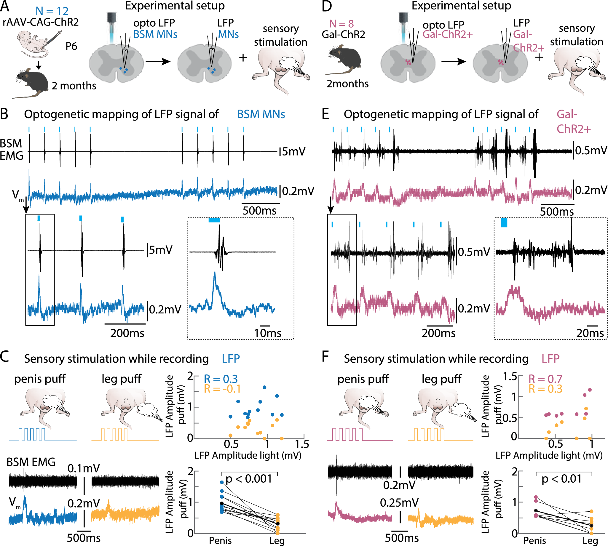
Here we mapped the spinal circuit controlling the bulbospongiosus muscle, essential for sperm expulsion in mice. Our findings show that bulbospongiosus muscle-motor neurons receive input from galanin-expressing neurons, which integrate genital sensory signals.
Stimulating these neurons induces bulbospongiosus activity, but responses vary with spinalization, internal state, and decrease with repeated stimulation.
Ablating galanin-positive neurons altered ejaculation latency and copulatory patterns. These results suggest that spinal circuits influence not only ejaculation but also copulation and arousal, challenging the traditional view of spinal control in copulation."

More than a reflex: How the spine shapes sex


More than a reflex: how the spine shapes sex (original news release) "For decades, it was thought that while the brain orchestrated male sexual behaviour – arousal, courtship, and copulation – the spinal cord merely executed the final act: ejaculation. But a study from the Champalimaud Foundation (CF) challenges that tidy division. It reveals that a key spinal circuit is not only involved in ejaculation but also in arousal and shaping the choreography of sex, adding a surprising new dimension to our understanding of sexual behaviour in mammals."


Fig. 3: Gal+ neurons and BSM-MNs receive sensory input from the penis.