Showing posts with label transcriptomics. Show all posts

Wednesday, October 08, 2025

Data-driven fine-grained region discovery in the mouse brain with transformers

Amazing stuff!

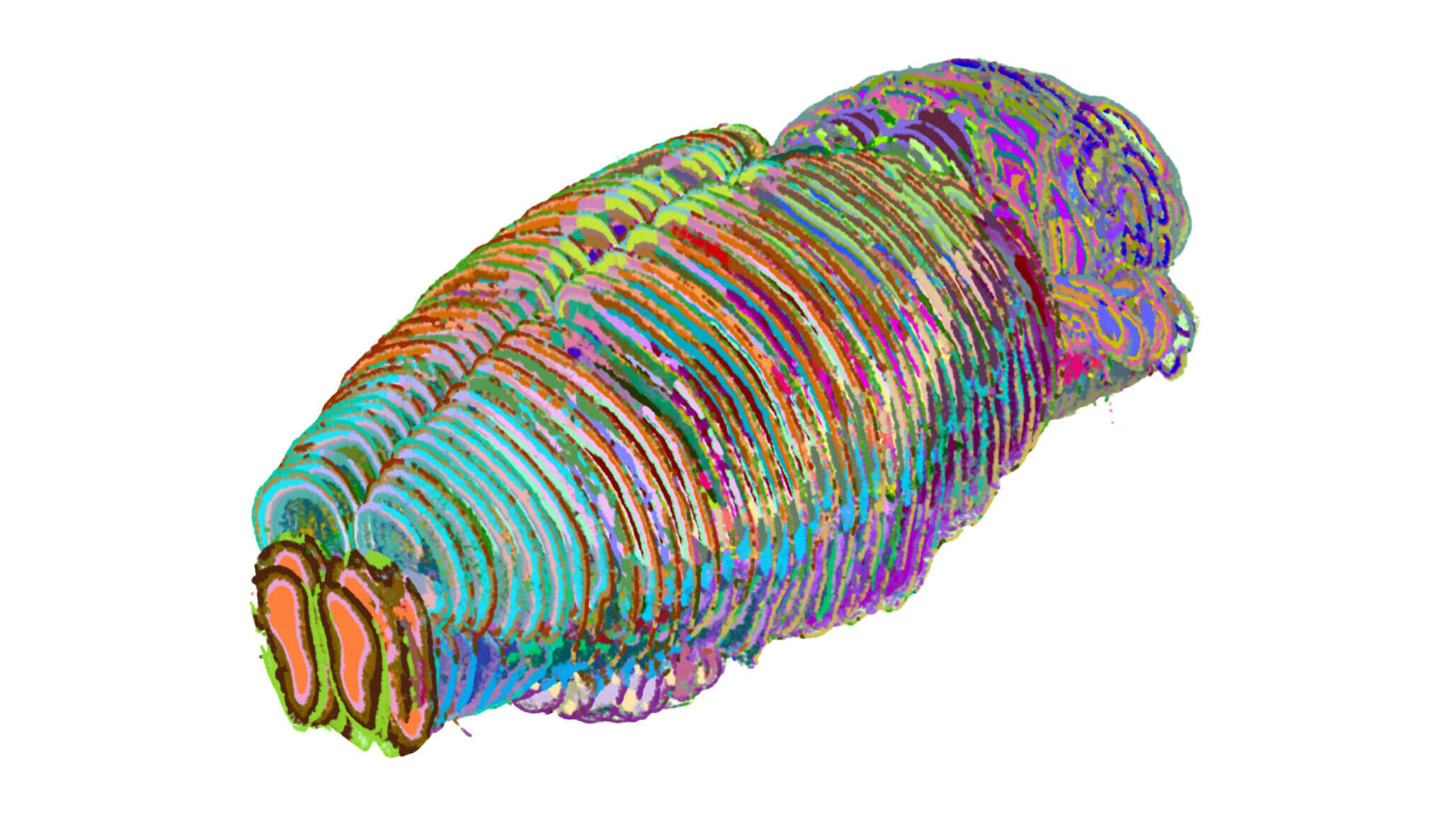
"Unleash artificial intelligence on an atlas that catalogs all cell types in a mouse brain, and—voilà! – you get one of the most granular and complex data-driven brain visualizations of any animal to date: a technicolor dream highlighting 1300 brain regions and subregions, including many that were previously uncharted.

“It’s like going from a map showing only continents and countries to one showing states and cities,” ... She and her colleagues developed a new AI tool called CellTransformer to make sense of vast spatial transcriptomics datasets that record where millions of cells sit and which genes they express. Traditional methods for mapping the brain’s cellular geography rely on hand-drawn, or annotated, atlases; CellTransformer’s algorithms infer from transcription patterns how cells are organized into functional neighborhoods.

When the researchers applied CellTransformer to the Allen Brain Cell Atlas, which encompasses nearly four million cells, it recapitulated known structures in the mouse brain such as the hippocampus but also uncovered hundreds of previously unannotated microregions, the team reported. ..."

"... CellTransformer, a powerful AI model that can automatically identify important subregions of the brain from massive spatial transcriptomics datasets. Spatial transcriptomics reveals where certain brain cell types are positioned in the brain but does not reveal regions of the brain based on their composition. Now, CellTransformer allows scientists to define brain regions and subdivisions based on calculations of shared cellular neighborhoods, much like sketching a city’s borders based on the types of buildings within it. ..."

From the abstract:
"Spatial transcriptomics offers unique opportunities to define the spatial organization of tissues and organs, such as the mouse brain. We address a key bottleneck in the analysis of organ-scale spatial transcriptomic data by establishing a workflow for self-supervised spatial domain detection that is scalable to multimillion-cell datasets.
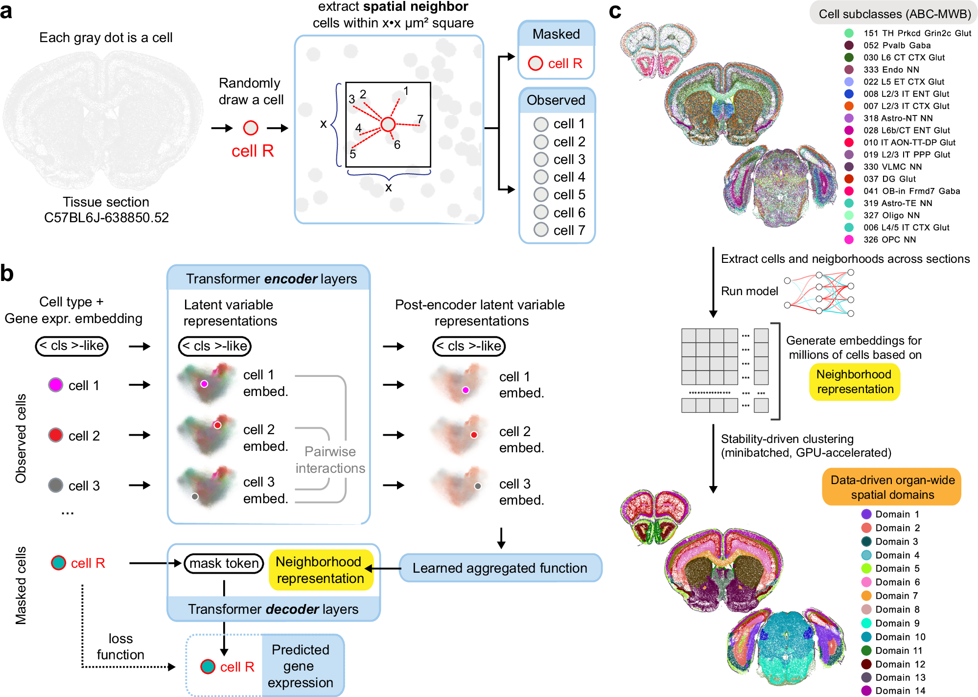
This workflow uses a self-supervised framework for learning latent representations of tissue spatial domains or niches. We use an encoder-decoder architecture, which we named CellTransformer, to hierarchically learn higher-order tissue features from lower-level cellular and molecular statistical patterns.
Coupling our representation learning workflow with minibatched GPU-accelerated clustering algorithms allows us to scale to multi-million cell MERFISH datasets where other methods cannot.
CellTransformer is effective at integrating cells across tissue sections, identifying domains highly similar to ones in existing ontologies such as Allen Mouse Brain Common Coordinate Framework (CCF) while allowing discovery of hundreds of uncataloged areas with minimal loss of domain spatial coherence. CellTransformer domains recapitulate previous neuroanatomical studies of areas in the subiculum and superior colliculus and characterize putatively uncataloged subregions in subcortical areas, which currently lack subregion annotation.
CellTransformer is also capable of domain discovery in whole-brain Slide-seqV2 datasets.
Our workflows enable complex multi-animal analyses, achieving nearly perfect consistency of up to 100 spatial domains in a dataset of four individual mice with nine million cells across more than 200 tissue sections.
CellTransformer advances the state of the art for spatial transcriptomics by providing a performant solution for the detection of fine-grained tissue domains from spatial transcriptomics data."

ScienceAdviser


Scientists create ChatGPT-like AI model for neuroscience to build detailed mouse brain map (original news release) "Artificial intelligence reveals undiscovered regions of the brain from large-scale spatial transcriptomics data"


Fig. 1: Overall training and architectural scheme for CellTransformer.

Three-dimensional representation of regions/subregions in mouse brain map created by CellTransformer. Fewer regions are generated for visual clarity and simplicity.

New technique offers bigger and better window into RNA activity in complex tissue

Amazing stuff!

"For the first time, scientists can view RNA molecules directly inside cells and tissue in minute detail and across the entire human genome concurrently, thanks to new technology created by a Yale research team.

The technique, known as Reverse-padlock Amplicon Encoding Fluorescence In Situ Hybridization (RAEFISH), solves a trade-off that researchers have long been forced to make: detail versus scope. Earlier tools required researchers to choose between seeing either a limited number of genes in high detail or seeing many genes but with a limited level of detail regarding their transcripts’ (RNAs’) location and how they interacted. ..."

From the highlights and abstract:
"Highlights
• RAEFISH enables genome-scale spatial transcriptome imaging with high resolution
• An efficient and robust probing and readout scheme that covers long and short transcripts
• RAEFISH maps spatially dependent transcriptomes in diverse cell and tissue contexts
• RAEFISH enables direct readout of gRNAs in image-based, high-content CRISPR screens

Summary
Recent breakthroughs in spatial transcriptomics technologies have enhanced our understanding of diverse cellular identities, spatial organizations, and functions. Yet existing spatial transcriptomics tools are still limited in either transcriptomic coverage or spatial resolution, hindering unbiased, hypothesis-free transcriptomic analyses at high spatial resolution.
Here, we develop reverse-padlock amplicon-encoding fluorescence in situ hybridization (RAEFISH), an image-based spatial transcriptomics method with whole-genome coverage and single-molecule resolution in intact tissues.
We demonstrate the spatial profiling of transcripts from 23,000 human or 22,000 mouse genes in single cells and tissue sections. 
Our analyses reveal transcript-specific subcellular localization, cell-type-specific and cell-type-invariant zonation-dependent transcriptomes, and gene programs underlying preferential cell-cell interactions.
Finally, we further develop our technology for the direct spatial readout of guide RNAs (gRNAs) in an image-based, high-content CRISPR screen.
Overall, these developments offer a broadly applicable technology that enables high-coverage, high-resolution spatial profiling of both long and short, native and engineered RNAs in many biomedical contexts."

New technique offers bigger and better window into RNA activity in complex tissue | Yale News "Yale researchers have created a new tool that will enable researchers to better view how RNA molecules function in tissue space."


Figure 1 RAEFISH enables genome-wide spatial transcriptomic profiling at single-molecule resolution


Figure 5 RAEFISH uncovers characteristics of spatial transcriptomic architectures and cell-cell interactions in mouse placenta


Sunday, December 08, 2024

Mouse study captures and maps aging process of skeletal muscles at the cellular level and in great detail

Good news! It is always good to have a detailed atlas! 

These poor mice were subjected to snake venom.

"As muscles age, their cells lose the ability to regenerate and heal after injury. Cornell Engineering researchers have created the most comprehensive portrait to date of how that change, in mice, unfolds over time and across the complicated architecture of muscle tissue. ...

“Does the decline in regeneration seen in old muscles come from changes to the stem cells that drive the repair process themselves, or does it come from changes in the way that they are instructed by other cell types?” ...

researchers sampled cells from young, old and geriatric mice at six time points after inducing injury via a variant of snake venom toxin. They identified 29 defined cell types, including immune cells that exhibited differences in their abundance and reaction time between age groups, and muscle stem cells that self-renew in youth but stall out as muscles age. ...

The detailed assessment of many cell types over time showed discoordination in the process of muscle repair in older mice. Many immune cells, which coordinate tissue repair, show up at the wrong time. ..."

From the abstract:
"In aging, skeletal muscle regeneration declines due to alterations in both myogenic and non-myogenic cells and their interactions. This regenerative dysfunction is not understood comprehensively or with high spatiotemporal resolution. We collected an integrated atlas of 273,923 single-cell transcriptomes and high-resolution spatial transcriptomic maps from muscles of young, old and geriatric mice (~5, 20 and 26 months old) at multiple time points following myotoxin injury. We identified eight immune cell types that displayed accelerated or delayed dynamics by age. We observed muscle stem cell states and trajectories specific to old and geriatric muscles and evaluated their association with senescence by scoring experimentally derived and curated gene signatures in both single-cell and spatial transcriptomic data. This revealed an elevation of senescent-like muscle stem cell subsets within injury zones uniquely in aged muscles. This Resource provides a holistic portrait of the altered cellular states underlying muscle regenerative decline across mouse lifespan."

Mouse study captures aging process at the cellular level | Cornell Chronicle


Fig. 1: Assembly of scRNA-seq atlas of skeletal muscle regeneration across mouse aging.


Fig. 2: Age-related changes to cell dynamics during skeletal muscle regeneration.


Tuesday, August 30, 2022

Genetic clues to age-related macular degeneration revealed

Good news!

"Scientists ... reprogrammed stem cells to create models of diseased eye cells, and then analysed DNA, RNA and proteins to pinpoint the genetic clues. ...
The researchers took skin samples from 79 participants with and without the late stage of AMD, called geographic atrophy. Their skin cells were reprogrammed to revert to stem cells called induced pluripotent stem cells, and then guided with molecular signals to become retinal pigment epithelium cells, which are the cells affected in AMD. ...
Analysis of 127,600 cells revealed 439 molecular signatures associated with AMD, with 43 of those being potential new gene variants. Key pathways identified were subsequently tested within the cells and revealed differences in the energy-making mitochondria between healthy and AMD cells, rendering mitochondrial proteins as potential targets to prevent or alter the course of AMD. ..."

From the abstract:
"There are currently no treatments for geographic atrophy, the advanced form of age-related macular degeneration. ... Induced pluripotent stem cells generated from patients with geographic atrophy and healthy individuals were differentiated to retinal pigment epithelium. Integrating transcriptional profiles of 127,659 retinal pigment epithelium cells generated from 43 individuals with geographic atrophy and 36 controls with genotype data, we identify 445 expression quantitative trait loci in cis that are asssociated with disease status and specific to retinal pigment epithelium subpopulations. Transcriptomics and proteomics approaches identify molecular pathways significantly upregulated in geographic atrophy, including in mitochondrial functions, metabolic pathways and extracellular cellular matrix reorganization. Five significant protein quantitative trait loci that regulate protein expression in the retinal pigment epithelium and in geographic atrophy are identified - two of which share variants with cis- expression quantitative trait loci, including proteins involved in mitochondrial biology and neurodegeneration. Investigation of mitochondrial metabolism confirms mitochondrial dysfunction as a core constitutive difference of the retinal pigment epithelium from patients with geographic atrophy. This study uncovers important differences in retinal pigment epithelium homeostasis associated with geographic atrophy."

Genetic clues to age-related macular degeneration revealed | Garvan Institute of Medical Research The discovery of molecular signatures of age-related macular degeneration will help with better diagnosis and treatment of this progressive eye disease.


Fig. 1: Generation of RPE from iPSCs, identification, and characterization of RPE subpopulations


Monday, March 15, 2021

A deep dive into cells’ RNA transcriptomics

Amazing stuff! Discovering the secrets of life!

"... has developed a new RNA detection method named BOLORAMIS (short for “Barcoded Oligonucleotides Ligated On RNA Amplified for Multiplexed and parallel In Situ analyses”) that overcomes this problem. BOLORAMIS enables the design and uses a new type of DNA probe that directly binds its RNA target and allows the straight-forward synthesis of a barcoded DNA amplicon, which can be visualized by fluorescent in situ hybridization (FISH) or sequenced in situ. BOLORAMIS enables the analysis of different classes of RNAs with higher specificity and sensitivity than FISSEQ and other methods, works in the context of cells and tissues, and can be highly multiplexed. ...
“We used a co-culture system of neuronal cells and brain microglia which are known to interact in many normal and disease processes, and targeted 96 different messenger RNAs simultaneously,” ... “This allowed us to uncover the spatial relationships between specific cells and RNAs.” ...
“In future research, BOLORAMIS’s high functionality in complex human tissues and human-specific organoids may well give us an edge in deciphering RNA signatures related to neurological disorders,” ...
With BOLORAMIS we have solved some of the challenges that technologies in the spatial transcriptomics field are facing. It gives us a significant advantage for understanding the behavior of molecular networks in normal and pathological processes, and for investigating new drug targets, and developing clinical diagnostics in the native context of tissues ..."


"We present barcoded oligonucleotides ligated on RNA amplified for multiplexed and parallel insitu analyses (BOLORAMIS), a reverse transcription-free method for spatially-resolved, targeted, in situ RNA identification of single or multiple targets. ..."

A deep dive into cells’ RNA reality New highly sensitive, specific, and multiplexable RNA detection method advances in situ transcriptomics with potential for a range of biomedical applications

Here is the link to the underlying paper: